17.2 Cerebral palsy and neurodegenerative disorders
Cerebral palsy
Cerebral palsy (CP) describes a group of permanent disorders of the development of movement and posture, causing activity limitation, that are attributed to non-progressive disturbances that occurred in the developing fetal or infant brain. The motor disorders of cerebral palsy are often accompanied by disturbances of sensation, perception, cognition, communication, and behaviour, by epilepsy, and by secondary musculoskeletal problems.
(Rosenbaum P et al 2007 Dev Med Child Neurol Suppl 109:8–14)
The term is generally applied to children with permanent motor impairment due to non-progressive brain disorders occurring before the age of 5 years. There are many different causes, a wide range of manifestations of the motor disorder, and various associated problems.
Cerebral palsy is not a single disorder, but a group of disorders with diverse implications for children and their families. For some young people with mild cerebral palsy, the only motor deficit may be a minimal hemiplegia, causing clumsiness with certain movements. In other children with severe cerebral palsy, the motor deficit may be spastic quadriplegia with little or no independent movement. Because each child with cerebral palsy is different, individual assessment and treatment are essential.
Prevalence
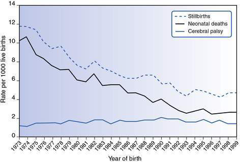
Cerebral palsy is the most common physical disability in childhood. The prevalence of cerebral palsy is between 2.0 and 2.5 per 1000 live births and has remained fairly stable since 1970 (Fig. 17.2.1).
![image](/wp-content/uploads/2016/08/B9780702042928000554_f17-04-9780702042928.jpg)
Fig. 17.2.1 Cerebral palsy, stillbirth and neonatal death rates per 1000 births in Victoria using published and unpublished data from the Victorian Cerebral Palsy Register and the Victorian Perinatal Data Collection Unit.
(Source: Reid S, Lanigan A, Reddihough DS 2005 Report of the Victorian Cerebral Palsy Register.)
Aetiology
The cause of cerebral palsy is unknown in many children. Known risk factors include low birth weight, prematurity and multiple pregnancy. In a significant proportion of children who have cerebral palsy, there appears to have been no single event but rather a sequence of events or ‘causal pathways’ that have culminated in the motor damage.
Historical aspects
There has been a fundamental change in our understanding of aetiological factors during the past 20 years. Before this, most cases of cerebral palsy were thought to be caused by lack of oxygen during labour or at birth, and it was expected that improvement in obstetrics and neonatal care would lower cerebral palsy rates. However, despite an increased use of interventions such as caesarean section and electronic fetal monitoring, cerebral palsy rates have remained constant.
Current research suggests that about 8–10% of cases are associated with perinatal asphyxia, a condition in which there have been perinatal events likely to reduce oxygen supply, evidenced by significant acidosis, and failure of function in at least two organs (usually the brain and kidney). Perinatal asphyxia may not necessarily be the primary cause of the cerebral palsy and is generally not preventable. Because it is often impossible to ascribe clinical signs and symptoms to an event during birth, the term ‘birth asphyxia’ should be avoided.
Current knowledge about aetiology
When does the brain insult occur?
• Prenatal events are responsible for approximately 75% of all cases of cerebral palsy.
• Perinatal events contribute 10–15%.
• Postneonatal causes (occurring after 28 days of life) account for about 10% of all cases.
A prenatal cause is assumed in the absence of clear evidence for a perinatal or postnatal cause.
What are the prenatal causes?
Malformations
Disturbances of brain development, usually between 12 and 20 weeks’ gestation, resulting in a variety of abnormalities. They may be identified by brain imaging, magnetic resonance imaging (MRI) being the preferred investigation. Some malformations have a genetic basis.
Infective
Maternal infections during the first and second trimesters of pregnancy, including the TORCH group of organisms (toxoplasmosis, rubella, cytomegalovirus and herpes simplex virus). There is also evidence that maternal infections in the perinatal period may form part of the causal pathway to cerebral palsy.
Why are premature and low-birth-weight infants at risk of cerebral palsy?
Premature and low-birth-weight children have a higher risk of cerebral palsy. For those born at less than 33 weeks’ gestation, the risk is up to 30 times higher than for those born at term. Some premature infants develop brain damage from complications of their immaturity, such as intraventricular haemorrhage, whereas others are damaged earlier in pregnancy. Intrauterine growth retardation is associated with cerebral palsy in both term and pre-term infants.
Periventricular white matter injury and the more localized pattern of periventricular leukomalacia is a common radiological finding in premature children with cerebral palsy. It is caused by an ischaemic process, usually occurring between 28 and 34 weeks’ gestation, in the watershed zone that exists in the periventricular white matter of the immature brain. Periventricular white matter injury is also found in infants born at term, suggesting that the insult occurred early in the third trimester even though the pregnancy progressed to term.
Why is multiple pregnancy a risk factor?
Multiple pregnancy is associated with pre-term delivery, poor intrauterine growth, birth defects and intrapartum complications and with an increased risk of both mortality and cerebral palsy. Intrauterine death of a co-twin is associated with a substantially increased rate of cerebral palsy.
What are the postneonatal causes of cerebral palsy?
• Infections, for example, meningitis, encephalitis and septicaemia.
• Injuries may be accidental (such as motor vehicle accidents and near drowning episodes), or non-accidental. Improved road safety and mandatory fencing around home swimming pools are important preventative measures.
• Apparent life-threatening events and cerebrovascular accidents.
• Meningitis, septicaemia and infections such as malaria are important causes of cerebral palsy in developing countries.
Caitlin is aged 2 years and 6 months. Her mother went into labour at 33 weeks’ gestation after an uneventful pregnancy. The delivery was rapid. Apgar scores were 6 at 1 minute and 8 at 5 minutes. Her parents remember some panic in the labour ward and felt that more could have been done to slow the labour. Caitlin developed hyaline membrane disease and mild jaundice. In the early neonatal period she had difficulty sucking, which was attributed to her prematurity. She was slow in her motor development and did not sit until the age of 15 months. A diagnosis of cerebral palsy was made at that time.
When Caitlin was 2 years old, her parents requested an opinion as to whether subsequent children were likely to have cerebral palsy, believing that her prematurity and problems at birth were responsible for her condition. MRI of the brain demonstrated a brain malformation with bilateral clefts in the cerebral cortex, dating the problems to early pregnancy rather than the perinatal period.
• Cerebral palsy is a diverse disorder with multiple risk factors and aetiologies.
• When determining aetiology, distinguish risk factors from causes.
• Most cases of cerebral palsy relate to events long before birth.
• Perinatal asphyxia is responsible for only a small proportion of cases (approximately 8–10%).
• It is important to establish the cause of cerebral palsy if at all possible. It is helpful for families and essential for genetic counselling.
• Magnetic resonance imaging (MRI) should be undertaken if the cause is not apparent.
Classification
Cerebral palsy is classified by motor type, topographical distribution and the severity of the motor disorder.
Type of motor disorder
Cerebral palsy is a disorder of movement (difficulties with voluntary movement and/or abnormal movements), posture and muscle tone. Children with cerebral palsy have various types of movement disorder.
Spastic cerebral palsy (70%)
This is the most common type. Spasticity involves increased muscle tone with characteristic clasp-knife quality. Children with spasticity often have underlying weakness. In spastic cerebral palsy, there is damage to the motor cortex or corticospinal tracts, in contrast to dyskinetic and ataxic cerebral palsy, which are associated with abnormalities of the basal ganglia and cerebellum, respectively.
Dyskinetic cerebral palsy (10–15%)
This refers to a group of cerebral palsies with involuntary movements and is characterized by abnormalities of tone involving the whole body. Several terms are used within this group:
Topographical distribution
The terms diplegia, hemiplegia and quadriplegia generally apply to children with spastic cerebral palsy as the extrapyramidal types (dyskinesia and ataxia) usually involve four limbs:
• The term diplegia is used where the predominant problem is in the lower limbs but signs are usually also present in the upper limb. Most of these children have normal intelligence. Spastic diplegia is the pattern most commonly seen in premature infants who have the radiological finding of periventricular white matter injury.
• Children with spastic hemiplegia usually have normal intelligence, frequently have epilepsy (50–70%) and visual deficits (homonymous hemianopsia), and may have sensory impairments in the upper limb.
• Children with spastic quadriplegia often have intellectual disability, epilepsy and visual impairment. Poor trunk control and oromotor difficulties may also be present.
Severity of the motor disorder
The Gross Motor Function Classification System (GMFCS) provides information about the severity of the movement problems based on children’s motor abilities and their need for walking frames, wheelchairs and other mobility devices. There are five levels: children in levels I and II walk independently; children in level III need walking frames or elbow crutches; and children in levels IV and V use wheelchairs. This classification system does not consider cognitive and other deficits.
Growth Motor Development Curves for each of the GMFCS levels are available and provide some guidance regarding prognosis for motor development.
The Manual Abilities Classification System (MACS) describes how children with cerebral palsy use their hands to handle objects in daily activities. It also has five levels, from level I, where children handle objects easily and successfully, to level V, where children are unable to handle objects and have severely limited ability to perform even simple actions.
• Cerebral palsy can be classified according to motor type, distribution and severity.
• GMFCS provides information about severity of gross motor function; MACS provides information about how children use their hands.
• Classifying motor severity using the GMFCS provides information about motor prognosis.
• Co-morbidities such as epilepsy are more common in certain types of cerebral palsy.
![](https://freepngimg.com/download/social_media/63059-media-icons-telegram-twitter-blog-computer-social.png)
Stay updated, free articles. Join our Telegram channel
![](https://clinicalpub.com/wp-content/uploads/2023/09/256.png)
Full access? Get Clinical Tree
![](https://videdental.com/wp-content/uploads/2023/09/appstore.png)
![](https://videdental.com/wp-content/uploads/2023/09/google-play.png)