Central Nervous System Infections

Chapter 595 Central Nervous System Infections


Infection of the central nervous system (CNS) is the most common cause of fever associated with signs and symptoms of CNS disease in children. Many microorganisms can cause infection. Nonetheless, specific pathogens are identifiable and are influenced by the age and immune status of the host and the epidemiology of the pathogen. In general, viral infections of the CNS are much more common than bacterial infections, which, in turn, are more common than fungal and parasitic infections. Infections caused by rickettsiae (Rocky Mountain spotted fever, Ehrlichia) are relatively uncommon but assume important roles under certain epidemiologic circumstances. Mycoplasma spp. can also cause infections of the CNS, although their precise contribution is often difficult to determine.


Regardless of etiology, most patients with CNS infection have similar clinical manifestations. Common symptoms include headache, nausea, vomiting, anorexia, restlessness, altered state of consciousness, and irritability; most of these symptoms are nonspecific. Common signs of CNS infection, in addition to fever, include photophobia, neck pain and rigidity, obtundation, stupor, coma, seizures, and focal neurologic deficits. The severity and constellation of signs are determined by the specific pathogen, the host, and the area of the CNS affected.


Infection of the CNS may be diffuse or focal. Meningitis and encephalitis are examples of diffuse infection. Meningitis implies primary involvement of the meninges, whereas encephalitis indicates brain parenchymal involvement. Because these anatomic boundaries are often not distinct, many patients have evidence of both meningeal and parenchymal involvement and should be considered to have meningoencephalitis. Brain abscess is the best example of a focal infection of the CNS. The neurologic expression of this infection is determined by the site and extent of the abscess(es) (Chapter 596).


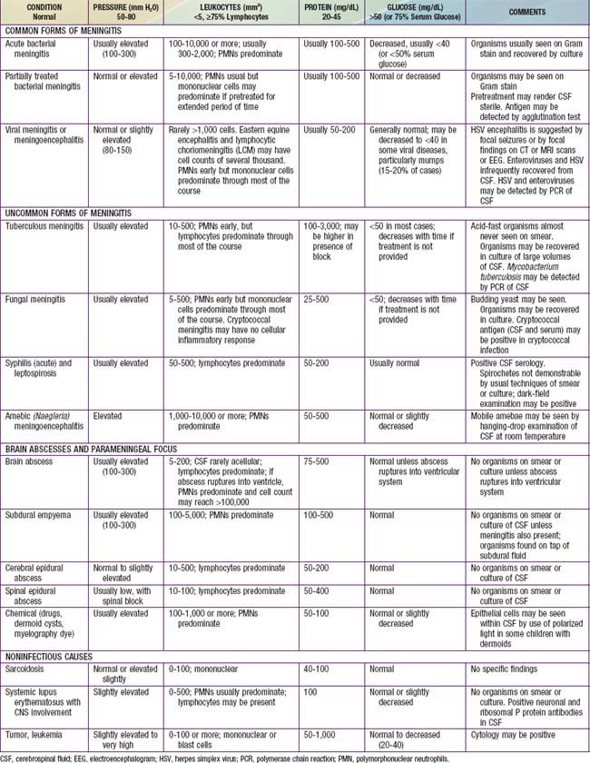
The diagnosis of diffuse CNS infections depends on examination of cerebrospinal fluid (CSF) obtained by lumbar puncture (LP). Table 595-1 provides an overview of the expected CSF abnormalities with various CNS disorders.


595.1 Acute Bacterial Meningitis Beyond the Neonatal Period


Bacterial meningitis is one of the most potentially serious infections occurring in infants and older children. This infection is associated with a high rate of acute complications and risk of long-term morbidity. The incidence of bacterial meningitis is sufficiently high in febrile infants that it should be included in the differential diagnosis of those with altered mental status and other evidence of neurologic dysfunction.


Epidemiology


A major risk factor for meningitis is the lack of immunity to specific pathogens associated with young age. Additional risks include recent colonization with pathogenic bacteria, close contact (household, daycare centers, college dormitories, military barracks) with individuals having invasive disease caused by N. meningitidis and H. influenzae type b, crowding, poverty, black or Native American race, and male gender. The mode of transmission is probably person-to-person contact through respiratory tract secretions or droplets. The risk of meningitis is increased among infants and young children with occult bacteremia; the odds ratio is greater for meningococcus (85 times) and H. influenzae type b (12 times) relative to that for pneumococcus.


Specific host defense defects due to altered immunoglobulin production in response to encapsulated pathogens may be responsible for the increased risk of bacterial meningitis in Native Americans and Eskimos. Defects of the complement system (C5-C8) have been associated with recurrent meningococcal infection, and defects of the properdin system have been associated with a significant risk of lethal meningococcal disease. Splenic dysfunction (sickle cell anemia) or asplenia (due to trauma, or congenital defect) is associated with an increased risk of pneumococcal, H. influenzae type b (to some extent), and, rarely, meningococcal sepsis and meningitis. T-lymphocyte defects (congenital or acquired by chemotherapy, AIDS, or malignancy) are associated with an increased risk of L. monocytogenes infections of the CNS.


Congenital or acquired CSF leak across a mucocutaneous barrier, such as cranial or midline facial defects (cribriform plate) and middle ear (stapedial foot plate) or inner ear fistulas (oval window, internal auditory canal, cochlear aqueduct), or CSF leakage through a rupture of the meninges due to a basal skull fracture into the cribriform plate or paranasal sinus, is associated with an increased risk of pneumococcal meningitis. The risk of bacterial meningitis, caused by S. pneumoniae, in children with cochlear implants, used for the treatment of hearing loss, is more than 30 times the risk in the general U.S. population. Lumbosacral dermal sinus and meningomyelocele are associated with staphylococcal and gram-negative enteric bacterial meningitis. CSF shunt infections increase the risk of meningitis due to staphylococci (especially coagulase-negative species) and other low virulence bacteria that typically colonize the skin.


Neisseria Meningitidis (Chapter 184)


Five serogroups of meningococcus, A, B, C, Y, and W-135, are responsible for disease. Meningococcal meningitis may be sporadic or may occur in epidemics. In the USA, serogroups B, C, and Y each account for ≈30% of cases, although serogroup distribution varies by location and time. Epidemic disease, especially in developing countries, is usually caused by serogroup A. Cases occur throughout the year but may be more common in the winter and spring and following influenza virus infections. Nasopharyngeal carriage of N. meningitidis occurs in 1-15% of adults. Colonization may last weeks to months; recent colonization places nonimmune younger children at greatest risk for meningitis. The incidence of disease occurring in association with an index case in the family is 1%, a rate that is 1,000-fold the risk in the general population. The risk of secondary cases occurring in contacts at daycare centers is about 1/1,000. Most infections of children are acquired from a contact in a daycare facility, a colonized adult family member, or an ill patient with meningococcal disease. Children younger than 5 yr have the highest rates of meningococcal infection. A 2nd peak in incidence occurs in persons between 15 and 24 yr of age. College freshmen living in dormitories have an increased incidence of infection compared to non–college-attending, age-matched controls.


The Centers for Disease Control and Prevention (CDC) recommends vaccination against meningococcus with 1 dose of a quadrivalent conjugate meningococcal vaccine between the ages of 11 and 18 yr and for persons 2-10 yr who are at increased risk for meningococcal disease. College freshmen living in dormitories who have not been previously vaccinated should also be vaccinated with MCV4.


Pathology and Pathophysiology


A meningeal purulent exudate of varying thickness may be distributed around the cerebral veins, venous sinuses, convexity of the brain, and cerebellum and in the sulci, sylvian fissures, basal cisterns, and spinal cord. Ventriculitis with bacteria and inflammatory cells in ventricular fluid may be present (more often in neonates), as may subdural effusions and, rarely, empyema. Perivascular inflammatory infiltrates also may be present, and the ependymal membrane may be disrupted. Vascular and parenchymal cerebral changes characterized by polymorphonuclear infiltrates extending to the subintimal region of the small arteries and veins, vasculitis, thrombosis of small cortical veins, occlusion of major venous sinuses, necrotizing arteritis producing subarachnoid hemorrhage, and, rarely, cerebral cortical necrosis in the absence of identifiable thrombosis have been described at autopsy. Cerebral infarction, resulting from vascular occlusion due to inflammation, vasospasm, and thrombosis, is a frequent sequela. Infarct size ranges from microscopic to involvement of an entire hemisphere.


Inflammation of spinal nerves and roots produces meningeal signs, and inflammation of the cranial nerves produces cranial neuropathies of optic, oculomotor, facial, and auditory nerves. Increased intracranial pressure (ICP) also produces oculomotor nerve palsy due to the presence of temporal lobe compression of the nerve during tentorial herniation. Abducens nerve palsy may be a nonlocalizing sign of elevated ICP.


Increased ICP is due to cell death (cytotoxic cerebral edema), cytokine-induced increased capillary vascular permeability (vasogenic cerebral edema), and, possibly, increased hydrostatic pressure (interstitial cerebral edema) after obstructed reabsorption of CSF in the arachnoid villus or obstruction of the flow of fluid from the ventricles. ICP may exceed 300 mm H2O; cerebral perfusion may be further compromised if the cerebral perfusion pressure (mean arterial pressure minus ICP) is <50 cm H2O due to systemic hypotension with reduced cerebral blood flow. The syndrome of inappropriate antidiuretic hormone secretion (SIADH) may produce excessive water retention and potentially increase the risk of elevated ICP (Chapter 553). Hypotonicity of brain extracellular spaces may cause cytotoxic edema after cell swelling and lysis. Tentorial, falx, or cerebellar herniation does not usually occur because the increased ICP is transmitted to the entire subarachnoid space and there is little structural displacement. Furthermore, if the fontanels are still patent, increased ICP is not always dissipated.


Hydrocephalus can occur as an acute complication of bacterial meningitis. It most often takes the form of a communicating hydrocephalus due to adhesive thickening of the arachnoid villi around the cisterns at the base of the brain. Thus, there is interference with the normal resorption of CSF. Less often, obstructive hydrocephalus develops after fibrosis and gliosis of the aqueduct of Sylvius or the foramina of Magendie and Luschka.


Raised CSF protein levels are due in part to increased vascular permeability of the blood-brain barrier and the loss of albumin-rich fluid from the capillaries and veins traversing the subdural space. Continued transudation may result in subdural effusions, usually found in the later phase of acute bacterial meningitis. Hypoglycorrhachia (reduced CSF glucose levels) is due to decreased glucose transport by the cerebral tissue.


Damage to the cerebral cortex may be due to the focal or diffuse effects of vascular occlusion (infarction, necrosis, lactic acidosis), hypoxia, bacterial invasion (cerebritis), toxic encephalopathy (bacterial toxins), elevated ICP, ventriculitis, and transudation (subdural effusions). These pathologic factors result in the clinical manifestations of impaired consciousness, seizures, cranial nerve deficits, motor and sensory deficits, and later psychomotor retardation.


Pathogenesis


Bacterial meningitis most commonly results from hematogenous dissemination of microorganisms from a distant site of infection; bacteremia usually precedes meningitis or occurs concomitantly. Bacterial colonization of the nasopharynx with a potentially pathogenic microorganism is the usual source of the bacteremia. There may be prolonged carriage of the colonizing organisms without disease or, more likely, rapid invasion after recent colonization. Prior or concurrent viral upper respiratory tract infection may enhance the pathogenicity of bacteria producing meningitis.


N. meningitidis and H. influenzae type b attach to mucosal epithelial cell receptors by pili. After attachment to epithelial cells, bacteria breach the mucosa and enter the circulation. N. meningitidis may be transported across the mucosal surface within a phagocytic vacuole after ingestion by the epithelial cell. Bacterial survival in the bloodstream is enhanced by large bacterial capsules that interfere with opsonic phagocytosis and are associated with increased virulence. Host-related developmental defects in bacterial opsonic phagocytosis also contribute to the bacteremia. In young, nonimmune hosts, the defect may be due to an absence of preformed IgM or IgG anticapsular antibodies, whereas in immunodeficient patients, various deficiencies of components of the complement or properdin system may interfere with effective opsonic phagocytosis. Splenic dysfunction may also reduce opsonic phagocytosis by the reticuloendothelial system.


Bacteria gain entry to the CSF through the choroid plexus of the lateral ventricles and the meninges and then circulate to the extracerebral CSF and subarachnoid space. Bacteria rapidly multiply because the CSF concentrations of complement and antibody are inadequate to contain bacterial proliferation. Chemotactic factors then incite a local inflammatory response characterized by polymorphonuclear cell infiltration. The presence of bacterial cell wall lipopolysaccharide (endotoxin) of gram-negative bacteria (H. influenzae type b, N. meningitidis) and of pneumococcal cell wall components (teichoic acid, peptidoglycan) stimulates a marked inflammatory response, with local production of tumor necrosis factor, interleukin 1, prostaglandin E, and other inflammatory mediators. The subsequent inflammatory response is characterized by neutrophilic infiltration, increased vascular permeability, alterations of the blood-brain barrier, and vascular thrombosis. Meningitis-associated brain injury is not simply caused by viable bacteria but occurs as a consequence of the host reaction to the inflammatory cascade initiated by bacterial components.


Rarely, meningitis may follow bacterial invasion from a contiguous focus of infection such as paranasal sinusitis, otitis media, mastoiditis, orbital cellulitis, or cranial or vertebral osteomyelitis or may occur after introduction of bacteria via penetrating cranial trauma, dermal sinus tracts, or meningomyeloceles.


Clinical Manifestations


The onset of acute meningitis has 2 predominant patterns. The more dramatic and, fortunately, less common presentation is sudden onset with rapidly progressive manifestations of shock, purpura, disseminated intravascular coagulation (DIC), and reduced levels of consciousness often resulting in progression to coma or death within 24 hr. More often, meningitis is preceded by several days of fever accompanied by upper respiratory tract or gastrointestinal symptoms, followed by nonspecific signs of CNS infection such as increasing lethargy and irritability.


The signs and symptoms of meningitis are related to the nonspecific findings associated with a systemic infection and to manifestations of meningeal irritation. Nonspecific findings include fever, anorexia and poor feeding, headache, symptoms of upper respiratory tract infection, myalgias, arthralgias, tachycardia, hypotension, and various cutaneous signs, such as petechiae, purpura, or an erythematous macular rash. Meningeal irritation is manifested as nuchal rigidity, back pain, Kernig sign (flexion of the hip 90 degrees with subsequent pain with extension of the leg), and Brudzinski sign (involuntary flexion of the knees and hips after passive flexion of the neck while supine). In some children, particularly in those younger than 12-18 mo, Kernig and Brudzinski signs are not consistently present. Indeed fever, headache, and nuchal rigidity are present in only 40% of adults with bacterial meningitis. Increased ICP is suggested by headache, emesis, bulging fontanel or diastasis (widening) of the sutures, oculomotor (anisocoria, ptosis) or abducens nerve paralysis, hypertension with bradycardia, apnea or hyperventilation, decorticate or decerebrate posturing, stupor, coma, or signs of herniation. Papilledema is uncommon in uncomplicated meningitis and should suggest a more chronic process, such as the presence of an intracranial abscess, subdural empyema, or occlusion of a dural venous sinus. Focal neurologic signs usually are due to vascular occlusion. Cranial neuropathies of the ocular, oculomotor, abducens, facial, and auditory nerves may also be due to focal inflammation. Overall, about 10-20% of children with bacterial meningitis have focal neurologic signs.


Seizures (focal or generalized) due to cerebritis, infarction, or electrolyte disturbances occur in 20-30% of patients with meningitis. Seizures that occur on presentation or within the 1st 4 days of onset are usually of no prognostic significance. Seizures that persist after the 4th day of illness and those that are difficult to treat may be associated with a poor prognosis.


Alterations of mental status are common among patients with meningitis and may be due to increased ICP, cerebritis, or hypotension; manifestations include irritability, lethargy, stupor, obtundation, and coma. Comatose patients have a poor prognosis. Additional manifestations of meningitis include photophobia and tache cérébrale, which is elicited by stroking the skin with a blunt object and observing a raised red streak within 30-60 sec.


Diagnosis


The diagnosis of acute pyogenic meningitis is confirmed by analysis of the CSF, which typically reveals microorganisms on Gram stain and culture, a neutrophilic pleocytosis, elevated protein, and reduced glucose concentrations (see Table 595-1). LP should be performed when bacterial meningitis is suspected. Contraindications for an immediate LP include (1) evidence of increased ICP (other than a bulging fontanel), such as 3rd or 6th cranial nerve palsy with a depressed level of consciousness, or hypertension and bradycardia with respiratory abnormalities (Chapter 584); (2) severe cardiopulmonary compromise requiring prompt resuscitative measures for shock or in patients in whom positioning for the LP would further compromise cardiopulmonary function; and (3) infection of the skin overlying the site of the LP. Thrombocytopenia is a relative contraindication for LP. If an LP is delayed, empirical antibiotic therapy should be initiated. CT scanning for evidence of a brain abscess or increased ICP should not delay therapy. LP may be performed after increased ICP has been treated or a brain abscess has been excluded.


Blood cultures should be performed in all patients with suspected meningitis. Blood cultures reveal the responsible bacteria in up to 80-90% of cases of meningitis.


Lumbar Puncture (Chapter 584)


The CSF leukocyte count in bacterial meningitis usually is elevated to >1,000/mm3 and, typically, there is a neutrophilic predominance (75-95%). Turbid CSF is present when the CSF leukocyte count exceeds 200-400/mm3. Normal healthy neonates may have as many as 30 leukocytes/mm3 (usually <10), but older children without viral or bacterial meningitis have <5 leukocytes/mm3 in the CSF; in both age groups there is a predominance of lymphocytes or monocytes.


A CSF leukocyte count <250/mm3 may be present in as many as 20% of patients with acute bacterial meningitis; pleocytosis may be absent in patients with severe overwhelming sepsis and meningitis and is a poor prognostic sign. Pleocytosis with a lymphocyte predominance may be present during the early stage of acute bacterial meningitis; conversely, neutrophilic pleocytosis may be present in patients in the early stages of acute viral meningitis. The shift to lymphocytic-monocytic predominance in viral meningitis invariably occurs within 8 to 24 hr of the initial LP. The Gram stain is positive in 70-90% of patients with untreated bacterial meningitis.


A diagnostic conundrum in the evaluation of children with suspected bacterial meningitis is the analysis of CSF obtained from children already receiving antibiotic (usually oral) therapy. This is an important issue, because 25-50% of children being evaluated for bacterial meningitis are receiving oral antibiotics when their CSF is obtained. CSF obtained from children with bacterial meningitis, after the initiation of antibiotics, may be negative on Gram stain and culture. Pleocytosis with a predominance of neutrophils, elevated protein level, and a reduced concentration of CSF glucose usually persist for several days after the administration of appropriate intravenous antibiotics. Therefore, despite negative cultures, the presumptive diagnosis of bacterial meningitis can be made. Some clinicians test CSF for the presence of bacterial antigens if the child has been pretreated with antibiotics and the diagnosis of bacterial meningitis is in doubt. These tests have technical limitations.


A traumatic LP may complicate the diagnosis of meningitis. Repeat LP at a higher interspace may produce less hemorrhagic fluid, but this fluid usually also contains red blood cells. Interpretation of CSF leukocytes and protein concentration are affected by LPs that are traumatic, although the Gram stain, culture, and glucose level may not be influenced. Although methods for correcting for the presence of red blood cells have been proposed, it is prudent to rely on the bacteriologic results rather than attempt to interpret the CSF leukocyte and protein results of a traumatic LP.


Differential Diagnosis


In addition to S. pneumoniae, N. meningitidis, and H. influenzae type b, many other microorganisms can cause generalized infection of the CNS with similar clinical manifestations. These organisms include less typical bacteria, such as Mycobacterium tuberculosis, Nocardia spp., Treponema pallidum (syphilis), and Borrelia burgdorferi (Lyme disease); fungi, such as those endemic to specific geographic areas (Coccidioides, Histoplasma, and Blastomyces) and those responsible for infections in compromised hosts (Candida, Cryptococcus, and Aspergillus); parasites, such as Toxoplasma gondii and those that cause cysticercosis and, most frequently, viruses (Chapter 595.2) (Table 595-2). Focal infections of the CNS including brain abscess and parameningeal abscess (subdural empyema, cranial and spinal epidural abscess) may also be confused with meningitis. In addition, noninfectious illnesses can cause generalized inflammation of the CNS. Relative to infections, these disorders are uncommon and include malignancy, collagen vascular syndromes, and exposure to toxins (see Table 595-2).


Table 595-2 CLINICAL CONDITIONS AND INFECTIOUS AGENTS ASSOCIATED WITH ASEPTIC MENINGITIS


VIRUSES


BACTERIA


BACTERIAL PARAMENINGEAL FOCUS


FUNGI


PARASITES (EOSINOPHILIC)


PARASITES (NONEOSINOPHILIC)


POSTINFECTIOUS


SYSTEMIC OR IMMUNOLOGICALLY MEDIATED


Stay updated, free articles. Join our Telegram channel

Jun 18, 2016 | Posted by in PEDIATRICS | Comments Off on Central Nervous System Infections

Full access? Get Clinical Tree

Get Clinical Tree app for offline access