Author (year)
Incidence in reproductive age group (%)
Number of cases
Maternal mortality (%)
Foetal mortality (%)
Bhattt (1974) [1]
NA
28
71.4
82.1
Jain (1993) [2]
13.3
25
20
36
Akhtar(1994) [3]
7.1
50
70
72
Sarkar(1996) [4]
NA
20
0
60
Prassana (1996) [5]
15
6
16.6
16.6
Gaffar (2007) [6]
14.9
32
71.8
81.2
Zalquarnain (2012) [7]
12.7
87
21.8
54.02
Aggarwal (2014) [8]
12.2
49
67.3
69.3
The paucity of data has made it difficult to frame the standard management guidelines of such patients. This chapter aims to describe the management of pregnant female with thermal injuries on the basis of limited available literature.
Classification and Estimation of Burn Wounds
Burn Depth
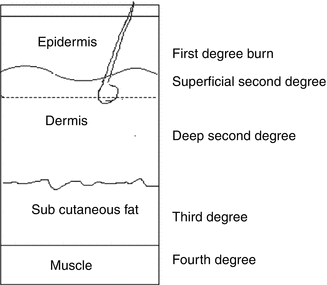
It is classified on the basis of degree of injury to the epidermis, dermis, subcutaneous fat and underlying structures.
First-degree burns are confined to the epidermis. These burns are painful, erythematous and blanch to the touch with an intact epidermal barrier. These burns do not result in scarring.
Second-degree burns are divided into two types: superficial and deep. This division is based on the depth of injury into the dermis. Superficial dermal burns are erythematous, painful and blanch to touch and often lead to blister formation. These wounds re-epithelialize spontaneously from retained epidermal structures in the rete ridges, hair follicles and sweat glands in 7–14 days. Deep dermal burns that extend into the reticular dermis, appear more pale and mottled, and do not blanch to touch but remain painful to pinprick. These burns heal in 14–35 days by re-epithelialization from hair follicles and sweat gland keratinocytes, often with severe scarring.
Third-degree burns are full thickness through the epidermis and dermis and are characterized by a hard, leathery eschar that is painless and black, white or cherry red. These wounds heal by re-epithelialization from the wound edges. Deep dermal and full-thickness burns usually require excision with skin grafting.
Fourth-degree burns involve other organs beneath the skin, such as the muscle and bone (Fig. 26.1).

Fig. 26.1
Burn depth
Burn Size
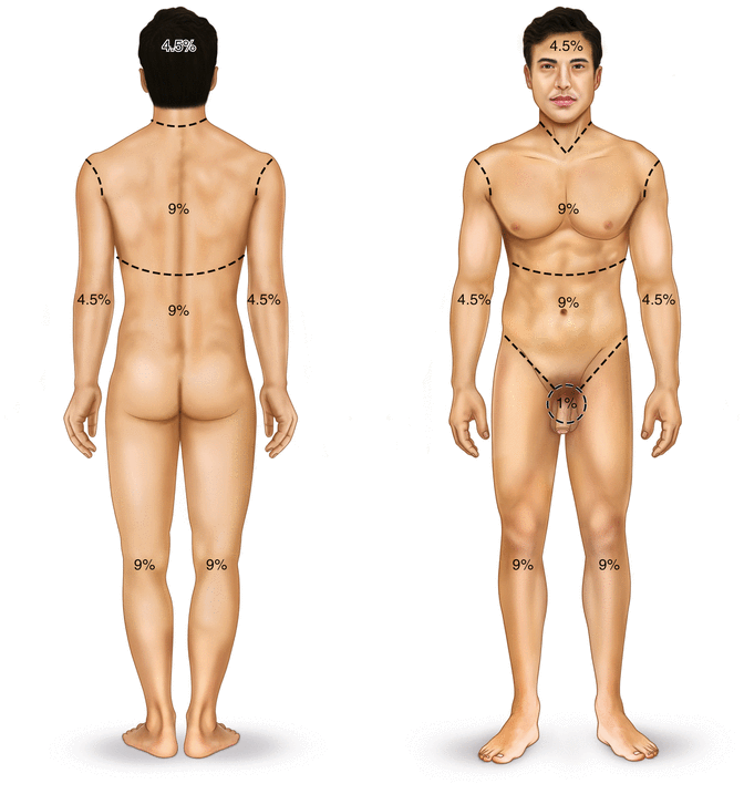
Burn size is generally assessed by the ‘rule of nines’ (Fig. 26.2). In adults, each upper extremity and the head and neck are 9 % of the total body surface area (TBSA), the lower extremities and the anterior and posterior trunk are 18 % each, and the perineum and genitalia are considered to be 1 % of the TBSA. For smaller burns, the area of the open hand (including the palm and the extended fingers) of the patient is taken as 1 % TBSA.


Fig. 26.2
Burn size
Management
In addition to preterm labour and intrauterine foetal death, the pregnant woman with major burns is at risk of all the serious complications that can occur in a nonpregnant woman like cardiovascular instability, respiratory distress, sepsis, liver failure and renal failure. Therefore, a multidisciplinary approach involving a plastic surgeon, obstetrician and intensivist is required for the management of burned pregnant patient. Going by the high incidence of burns in reproductive age group (up to 15 %), all female burned patients of child-bearing age should be tested for pregnancy unless the pregnancy is obvious. Early diagnosis of pregnancy helps in avoiding teratogenic drugs and ionizing imaging studies and at the same time helps in initiating optimal therapy for better outcome [9].
Fluid Therapy
Proper fluid management is critical to survival following major thermal injury. One of the principal causes of multiple organ failure system (MOFS) is found to be inadequate fluid resuscitation. The primary goal of fluid resuscitation is to ensure end-organ perfusion by replacing fluid that is sequestrated as a result of the thermal injury. Pregnancy is associated with hyperdynamic circulation. As pregnancy progresses, there is also progressive fall in colloid osmotic pressure. These changes, along with an increase capillary permeability in burned patient, predispose the burned pregnant patient to additional fluid loss beyond the amount as seen in nonpregnant individuals. Because uterus has little capacity for autoregulation, early and aggressive fluid resuscitation is vital to avoid maternal hypotension and resultant utero-placental hypoperfusion and foetal hypoxia in these patients [10, 11].
In nonpregnant patients, the fluid requirement in early postburn is estimated using Parkland formula. According to this, the fluid requirement in the first 24-h postburn period is 4 ml/kg body weight per percent of burned body surface area. One half of this fluid is to be given in the first 8 h and the rest in the next 16 h. However, it has been found that this formula underestimates the fluid requirement in pregnant patients by almost 50 %. Therefore, it is suggested that volume resuscitation should be titrated to other parameters like urine output (30–60 ml/h), mean arterial pressure, central venous pressure (CVP) and maternal and foetal heart rate, rather than by Parkland formula. As far as the choice of fluid is concerned, crystalloid, i.e. lactated Ringer solution, should be preferred as normal saline invariably leads to hyperchloremic metabolic acidosis [12].
Ventilation
Thermal injury to the respiratory tract is usually manifests as mucosal and submucosal erythema, oedema and ulceration. It is usually limited to upper airway because of reflex closure of vocal cords to hot air. The presence of significant intraoral and pharyngeal injury is an indication for early endotracheal intubation.
Supplemental oxygen and nursing in semi-sitting posture is recommended for all patients even in the absence of smoke inhalation injury. This improves tissue oxygenation, especially in third trimester when there is reduced functional residual capacity and end-expiratory volume. Inhalation injury must be suspected in patients who sustain burns in closed space or have facial burns. Inhaled carbon monoxide crosses placental barrier, binds to foetal Hb and can result in foetal hypoxemia. Supplementing 100% oxygen helps in reversing this by reducing half-life of carboxyhemoglobin. Ventilatory support should only be initiated when maternal pO2 is less than 60 mmHg [13].
Wound Management
Burn Wound Excision
For deeper wounds (deep partial thickness and full thickness), rather than waiting for spontaneous healing, the eschar is surgically removed and wound is closed by split skin graft or by flap. This approach is known as early excision and grafting (E&G). Any burn wound projected to take longer than 3 weeks to heal is a candidate for E&G within the first postburn week. Early tangential excision of burn wound and grafting improves maternal and foetal outcome by reducing septic complications. Removal of burn eschar also reduces the circulating level of prostaglandin E2 (PGE2) and a burn toxin-A lipoprotein complex released form damaged cell membranes, which are responsible for uterine stimulation [5, 14]. Early excision and grafting also minimizes excessive scar formation which is associated with secondary healing of wounds. Wounds over the abdomen and breasts should be treated first. This helps in pain-free stretching of abdominal wall as pregnancy develops, better supervision of growing foetus and easy performance of caesarean section if need arises. Better healing of breast wound prevents infection and sloughing of nipples and subsequent breastfeed. Early excision is recommended not only in patients who sustain minor burns but also in patients who has full-thickness major burns ranging from 25 to 65 % [10, 15].
Stay updated, free articles. Join our Telegram channel

Full access? Get Clinical Tree


