Parameter
Percentage of change
Cardiac output
40–50 %
Increase
Stroke volume
30 %
Increase
Heart rate
15–25 %
Increase
Intravascular volume
45 %
Increase
Systemic vascular resistance
20 %
Decrease
Systolic BP
Minimal
Diastolic BP
20 %
Decrease at mid-pregnancy
Pre-pregnant values at term
CVP
Unchanged
O2 consumption
30–40 %
Increase
Diagnosis and Evaluation of Heart Disease
Many women with heart disease would have been diagnosed and treated before pregnancy. Alternatively, heart disease may be diagnosed for the first time during pregnancy owing to symptoms precipitated by increased cardiac demands, or the patient might have tolerated heart disease well throughout the pregnancy and present during labour or postpartum period with acute symptoms for the first time, a not so unrare fact in India.
The classic symptoms of cardiac disease are palpitation, shortness of breath with exertion, and chest pain. Because these symptoms also may accompany normal pregnancy, a careful history and meticulous examination is needed to determine whether the symptoms are of particular concern in a patient with other reasons to suspect underlying cardiac disease.
A systolic murmur is present in 80 % of pregnant women, most likely due to the increased flow volume in the aorta and pulmonary artery. Any diastolic murmur and any systolic murmur that is loud (grade 3 or higher) or radiates to the carotids should be considered pathologic. Careful evaluation of jugular venous pulse, for peripheral cyanosis or clubbing and pulmonary crackles, is needed in women with suspected cardiac disease. The preferred next step evaluation includes transthoracic echocardiography but may not be available in an acute cardiac event. A chest X-ray is useful if CCF is suspected; dilated cardiac shadow with congestion in lungs with or without plural effusion may be seen. ECG may be helpful to suggest underlying heart disease, but both of these simple tests have limitation during pregnancy due to alteration in cardiac position due to gravid uterus late in pregnancy.
Ischaemic heart disease presenting for the first time during pregnancy is very challenging to diagnose. Heartburn due to GERD/oral iron intake and shortness of breath are not uncommon during pregnancy, and not only these but mild ECG changes may be normal findings during pregnancy besides elevated CPK-MB during pregnancy. Positive TROP-T test and change in cardiac enzyme (falling levels) will help to diagnose it retrogradely.
General Care
Deterioration in cardiac status during pregnancy is frequently insidious. Early registration and frequent and regular visits are a must. Attention is given to the HR, weight gain, and oxygen saturation. The physiological changes of pregnancy are usually continuous and offer adequate time for maternal compensation. Intercurrent events superimposed on pregnancy in the context of maternal heart disease are usually responsible for acute decompensation. The most common events are febrile episodes. Patients should be instructed to report to the hospital in case of fever, upper respiratory tract infection, and burning micturition. Iron and folic acid supplementation may decrease cardiac work.
First trimester dating scan would not only help in knowing exact EDD but also in monitoring growth of fetus in subsequent scan; detailed malformation scan would help if patient conceived on cardiac medication including fetal heart echocardiography specifically if mother is having congenital cardiac lesion.
Intrauterine growth restriction may be either due to ongoing medication or due to compromised oxygenation of fetus; Doppler studies may help in deciding pregnancy termination and mode of delivery.
Standard care in labour and delivery:
1.
Accurate diagnosis
2.
Close monitoring of maternal and fetal well-being
3.
Mode of delivery based on obstetric indications
4.
Prophylactic antibiotic when at risk for endocarditis, valvular heart disease, prosthetic heart valves, structured congenital heart diseases, previous infective endocarditis, and hypertrophic cardiomyopathy
5.
Maintenance of haemodynamic stability
6.
Avoidance of pain – epidural analgesia
7.
Avoidance of maternal pushing efforts – instrumental delivery
8.
Avoidance of maternal blood loss – active management of third stage of labour
Cardiac Lesions and the Associated Maternal Cardiac Risks during Pregnancy
Maternal mortality associated with heart disease in pregnancy
Group 1: mortality <1 % | |
Atrial septal defect Ventricular septal defect; PDA Pulmonary/tricuspid disease Tetralogy of Fallot, corrected; bioprosthetic valve Mitral stenosis, NYHA classes I and II | |
Group 2: mortality 5–15 % | |
2A | Mitral stenosis NYHA classes III–IV; aortic stenosis Coarctation of aorta, without valvular involvement Uncorrected tetralogy of Fallot Previous myocardial infarction Marfan syndrome with normal aorta Mitral stenosis with atrial fibrillation Artificial valve |
2B | |
Group 3: mortality 25–50 % | |
Primary pulmonary hypertension or Eisenmenger’s syndrome Coarctation of aorta, with valvular involvement Marfan syndrome with aortic involvement | |
NYHA (New York Heart Association) Functional Classification [4]
1.
Class I (mild) – no limitation of physical activity. Ordinary physical activity does not cause undue fatigue, palpitation, or dyspnoea.
2.
Class II (mild) – slight limitation of physical activity. Comfortable at rest but ordinary physical activity results in symptoms.
3.
Class III (moderate) – marked limitation of physical activity, comfortable at rest, but less than normal activity cause symptoms.
4.
Class IV (severe) – unable to carry out any physical activity without discomfort. Symptoms of cardiac insufficiency at rest.
Medical termination of pregnancy is to be advised to patients specifically with high risk of cardiac lesion, in view of increased mortality in such cases, but even low-risk patients can complicate in such an outcome.
Carpreg Score, Hamilton, and Thompson
Bad prognostic criteria will help in guiding outcome during this pregnancy (Table 5.1).
Table 5.1
Cardiac disease in pregnancy (CARPREG) risk score
One point for each: |
History of prior cardiac event or arrhythmias |
New York Heart Association functional classification > II or cyanosis |
Left heart obstruction (mitral valve area <2 cm2, aortic valve area <1.5 cm2 or left ventricular outflow tract gradient >30 mmHg) |
Left ventricular ejection fraction <0.40 |
Chance of cardiac complication: |
0 points = 5 % 1 point = 27 % ≥2 points = 75 % |
Valvular Disease
Mitral Stenosis
Mitral stenosis is the most common cardiac disease occurring mostly as a consequence of rheumatic heart disease. Risk of maternal complications in MS is strongly associated with the severity of MS, NYHA functional class, previous history of pulmonary oedema, and embolic phenomena. Complications include pulmonary oedema, right ventricular failure, and atrial arrhythmias with risk of embolisation. Pregnancy is detrimental to cardiac function in mitral stenosis for several reasons. Expanded blood volume can increase the risk of pulmonary congestion and oedema. The physiological tachycardia of pregnancy decreases filling time which leads to elevated left arterial pressure that causes pulmonary oedema and decreased forward flow that causes hypotension, fatigue, and syncope.
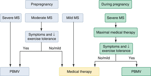
The severity of mitral stenosis is classified based on the valve area: a valve area of >1.5 cm2 is mild, 1.1–1.5 cm2 is moderate and <1 cm2 is severe. Treatment of mitral stenosis in patients who have a history of RHD includes daily prophylactic penicillin, gentle diuresis to prevent pulmonary oedema without decreasing placental function and ß-blockers as needed to prevent tachycardia. Atrial fibrillation can be treated with cardioversion; digoxin or ß-blockers may be used for rate control. Patients with atrial fibrillation should be anticoagulated to prevent systemic embolism. The most common surgical treatment of mitral stenosis is percutaneous balloon mitral valvotomy. This procedure should be preferably performed before conception but in women with critical mitral stenosis may be formed during second trimester of pregnancy with less fetal risk. [2]
Aortic Stenosis
Most common cause of AS in young women is a congenital bicuspid valve. RHD is a less common cause. The severity of AS can be described by average valve area or peak pressure gradient across the valve. Severe AS is defined as a peak gradient greater than 50 mmHg. Patients having severe AS have difficulty in achieving the increased cardiac output as their stroke volume is fixed by the obstructed valve, so heart rate is the key determinant of cardiac output. Bradycardia causes decreased cardiac output and hypotension; however, excessive tachycardia decreases excessive ventricular filling time and again causes decreased cardiac output and the risk for myocardial ischaemia. Women with severe AS are advised to undergo operative procedure before planning for pregnancy. Sudden death and irreversible heart failure are the most common causes of maternal death.
Stay updated, free articles. Join our Telegram channel

Full access? Get Clinical Tree


