Urine output is one of the key determinants of resuscitation. General urine output guidelines are for infants and children 2 cc per kilogram (kg) per hour, for adolescents 1 cc/kg/h, and for adults 0.5 cc/kg/h.
In terms of fluid resuscitation of the burned child, there are many guidelines published by various centers (Armour and Billmire 2009). Most will use crystalloid initially and then transition to some form of colloid.
It is critical to determine the need for transfer to a burn center. The American Burn Association has a specific list of criteria, which are as follows:
Partial thickness burns >10 % TBSA
Burns involving face, hands, feet, genitalia, perineum, or major joints
Third-degree burns (any age group)
Electrical burns (including lightning injury)
Chemical burns
Inhalation injury
Burns in patients with preexisting medical conditions
Burns and concomitant trauma where the burns pose the greater risk of morbidity/mortality
Burned children in hospital without qualified staff or equipment for care of child
Burn injury in patients who will need special social, emotional, or rehabilitative intervention
Even in the acute setting, the hand surgeon must be prepared for the chronic phase of care because burn care in children constantly requires planning for the next phase of care. This planning includes assessment of not only the actual deficits and defects but the potential ones that the reconstruction can create. By following these concepts, physicians can help to optimize recovery of the burn victim from the initial encounter. Failure to consider these issues may prolong recovery and reduce functional outcome.
It is critical that the hand specialist should be involved from the outset in the management of these patients. However, the hand specialist must also be aware of etiology of the injury and the real risk for concomitant trauma to other systems of the body. For that reason, the hand specialist must be familiar with the fundamental ATLS guidelines (see Advanced Trauma Life Support (ATLS) Student course manual 9th edition 2012).
Burn: Anatomy and Physiology
The hand is one of the most delicate instruments of the body. It is responsible for connecting to the outside world that includes work, touch, and communication. The anatomy of the hand is precise, and a detailed knowledge of the anatomy and function of the hand and its complex structures is essential for anyone charged with caring for these patients.
Assessment of Burn Depth
In the burn victim, an understanding of the anatomy and pathophysiology of burns is critical to patient management. Burns are characterized by the depth of injury into four categories (Table 1; Young 2006).
Table 1
Classification of burn injuries
Burn type | Depth | Skin changes | Healing |
|---|---|---|---|
First | Epidermis | Red coloration | Spontaneous in 3–5 days |
Second – superficial | Into upper dermis | Red, blistered, swollen | 7–14 days |
Second – deep | Into deep dermis | Red, swollen, blistered | Often requires intervention |
Third | Through deep dermis | White, charred, or translucent | Grafting or flap needed |
Fourth | Full thickness | Necrosis | Grafting or flap |
A first-degree burn is a superficial injury. The skin appears red and may be painful. The dermis is unaffected in these injuries. A sunburn is an example of a first-degree burn. These injuries require supportive care and will heal without scarring within 5–7 days (Fig. 1).
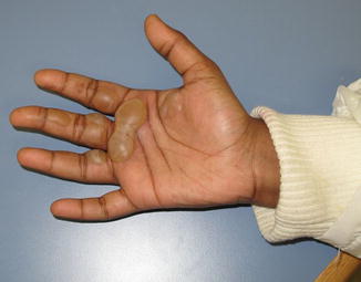
Fig. 1
Blistering of hand indicating superficial injury. Minimal care is needed beyond supportive measures (Courtesy of Shriners Hospital for Children, Philadelphia)
Second-degree burns can be superficial or deep (Fig. 2). Superficial second-degree burns are injuries to, but not through, the dermis. The skin is red, blistered, and swollen. Pain is prominent with a superficial second-degree burn. A superficial second-degree burn should heal spontaneously in 7–14 days. A deep second-degree burn is one that does not heal within 7–14 days. The dermis is injured in its full thickness and scarring will likely occur as a sequela.
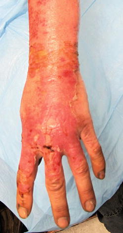
Fig. 2
Superficial injury . First-degree bordering on early second-degree injury. Note the redness akin to a bad sunburn. Treatment priorities are to return functional status as soon as possible. Silvadene was used to allow for a moist healing environment (Courtesy of M. Baumholtz, MD FACS)
Third-degree burns may appear whitish, charred, or translucent. Pinprick sensation is absent in the burned tissue because the nerve endings have been damaged by this injury, which traverses the dermis in its entirety. Because there are no dermal appendages to support wound healing, scar formation is inevitable and reconstruction using flaps or grafts is essential if the quantity of tissue loss is considerable.
Fourth-degree burns include full-thickness tissue loss to and including bone. Electrical injuries often cause fourth-degree burns. In these injuries, all overlying structures are dead and should be debrided.
As a general rule, all burns that are deep second degree, third degree, and fourth degree will require early excision and reconstruction. The nature of the reconstruction will depend on the location of the anatomic defect along with the functional deficit.
This section is concerned with the intersection of burn pathophysiology and hand function. To that end, the anatomy pertinent to caring for hand burns will be reviewed. Hand anatomy entails some unique features that will impact the manner in which burns are treated.
The volar, palmar skin of the hand provides padding for hand function with thick glabrous skin. However, there is relatively little room for swelling and expansion. This increases the risk for compartment syndromes.
The nail and nail bed are an often overlooked component of hand injuries, especially the burned hand. The nail and nail bed are specialized structures, which require both the matrices to make the nail and the proper support and surrounding tissues, which comprise the nail folds and distal phalanx.
The dorsal structures are more at risk due to the thin skin coverage and relatively little subcutaneous tissue. Specifically at risk is the PIP joint (Germann and Weigel 2011) due to its thin skin surface and the associated ligaments that arise from the extensor mechanism. Disruption of these structures can have devastating effect on function of the entire finger, even if the distal joint is uninvolved with the injury.
Physiology of Burn
A burn is divided into three areas of injury called zones. These three zones are described as:
Zone of coagulation
Zone of stasis
Zone of hyperemia
Irreversible tissue damage occurs in the zone of coagulation , while the zone of hyperemia will return to normal. The zone of stasis, however, is an area of tissue around the central coagulation zone, where the outcome can be modulated based on treatment. Proper resuscitation and care can salvage tissue in this zone, whereas inadequate resuscitation and treatment can lead to a worsening and loss of this tissue.
Mechanism of Thermal Injury
Heat injuries occur from the outside in and from superficial to deep. Injury is caused by flame or heat contact. The actual injury creates direct cellular destruction due to the thermal energy.
Cold injuries also occur from the outside in and from superficial to deep. However, at the cellular level there is cellular dehydration and crystal formation. Moreover, there is also the risk of cell damage that can occur during the rewarming phase as the crystals thaw and fluid shifts result in substantial edema.
Chemical burns can occur from acid, alkali, or specific chemicals common to hand burns. Knowledge of the causative agent is extremely valuable in treatment planning. The treatment for most chemical burns begins with copious irrigation. There are some exceptions to that. Hydrofluoric acid (HF) is one example. Exposure to HF often causes pronounced pain in the hands. This chemical is common in products used in auto detailing. Treatment may require either topical application or direct injection of calcium until the pain abates.
For management of several other specific chemical exposures, see Table 2.
Chemical agent | Treatment |
|---|---|
Chromic acid | Dilute sodium hyposulfite |
Sodium hypochlorite (common bleach) | Milk, egg white, starch |
Potassium permanganate (oxidizer) | Egg white |
Phenol (medication) | Mineral oil |
White phosphorous (smoke) | 2 % copper sulfate, oil |
Lye (degreaser) | Vinegar (acetic acid) or other weak acids such as lemon juice or orange juice |
Formaldehyde | Ammonium salts |
Mustard gas (chemical weapon) | Kerosene, gasoline, or mineral oil |
Hydrofluoric acid (wheel cleaner) | Calcium chloride |
Hydrochloric acid (swimming pool) | Soda lime, liquid soap |
Sulfuric acid (drain cleaner) | Magnesium oxide, liquid soap |
Elemental sodium | Mineral oil |
With regard to electrical injuries, the mechanism of damage continues to be debated. However, as would be expected, the majority of the damage is often the consequence of high-voltage energy passing through the tissues, especially the deeper structures. Whether it is due to the fact that the bone heats the surrounding tissue due to its resistance or that the electrical current flows through the deep muscle and nerves more easily, severe deep tissue injury can occur in the presence of seemingly innocuous superficial wounds.
Management of Acute Burns
Upper extremity burn injuries require long-term plans from their initial evaluation. Gone is the model of only excision and grafting for burns that are deep second degree and deeper. Instead, a carefully orchestrated plan that ultimately returns the patient to maximal function and minimal morbidity (both at the site of the burn and the donor sites) is essential.
The most important step is to stop the burn. This is done by extinguishing the fire and/or removing the offending agents and clothing. Placing the burned hand or finger under cool water can mitigate some of the secondary injury and can be comforting to the patient. However, as the wounds get larger, this becomes not only impractical but risks hypothermia in the burn patient. Therefore, one must be judicious in the use of large amounts of cooling.
Simple dry dressings are often the best first dressing. Keep the patient warm and use non-adherent dressings. This is especially true if the patient is to be transferred to a burn center where all the dressings will be removed as part of the initial survey. Once the wounds have been examined by the appropriate treating physicians, they can be dressed. Typical dressings in the acute period involve a non-adherent layer such as Adaptic®, Xeroform®, or Telfa® and often the application of some cream or ointment at the same time. A burn wound can be incredibly painful. For large areas or young children, dressing changes under sedation may be necessary.
Controversy exists regarding the use of systemic antibiotics for burn patients. In the initial period, there is little evidence to support the need for systemic antibiotics. Instead, centers mostly use a topical application based on depth and location of the burn. Below are some common choices for topical treatments (Palmieri and Greenhaigh 2002).
Common topical antimicrobials and their side effects:
Bacitracin ointment : This product has the advantage of being simple, cheap, and available. It has a broad spectrum of coverage, including Gram-positive and Gram-negative organisms. However, there is erythema following overuse that is not a true allergy but rather a reaction to a component in the product, which causes the skin to be red. While it is not an infection, it is often mistaken for one leading the patient or provider to use more and more ointment thinking that the ointment is treating the infection. Typically bacitracin is used for about 3–5 days after injury as the wounds heal. Subsequently, the patient may switch to using a bland entity such as Vaseline or A&D ointment. If the wound is worsening, it is necessary to change to a different treatment regimen.
Bactroban : This is an antimicrobial that is derived from pseudomonas species. It has good Gram-positive coverage and was originally intended for MRSA but resistance is emerging.
Silvadene : This is cheap and easy to apply. Its application is not painful. It is supplied as a white cream that takes on a yellowish tan color after being on the wound for a day or so. It provides good coverage for Gram-positive bacteria and good yeast coverage but demonstrates variable efficacy for Gram-negative organisms. There is a small risk of neutropenia and thrombocytopenia with prolonged use. Simple blood tests can monitor for this.
Sulfamylon: This is unique in that it provides good penetration of eschar. It is especially helpful for wounds near or involving cartilage because of this property. Sulfamylon provides coverage for both Gram-positive and Gram-negative organisms, but poor control of yeast. It is often prepared as an aqueous solution and patients will frequently feel pain during the application process. This property has led sulfamylon to be known as white lightening. Sulfamylon application can lead to metabolic acidosis because it inhibits carbonic anhydrase. For these reasons, it is not a first-line choice of treatment for most burn specialists.
Silver nitrate : This is an agent that predates all of the other agents described above. It stains tissue and clothing but provides good coverage for Gram-positive and Gram-negative organisms as well as yeast. Application is painless. The use of silver nitrate carries a remote risk of methemoglobinemia and argyria. Other more common risks include electrolyte abnormalities.
Nutrition and the Burn Patient
Nutritio n plays a major role in patients’ ability to heal and fight infection. Nutritional support is critical in management of the burn patient. Most calorie estimations of oral intake alone are grossly inadequate. Moreover, the formulas used for nutritional support are often insufficient in the face of burn and multiple trauma victims. It remains generally accepted that the patient’s own intestinal tract is the best route for nourishment whenever possible. Albumin and prealbumin are lab markers that can help guide nutritional support. The condition known as refeeding syndrome is exceedingly rare and many of the current-day fears about this process are unfounded. Nevertheless, all patients who can eat are encouraged to do so. The patient’s albumin and prealbumin are used to drive their nutritional support. A blood albumin level less than 2.7 g/dL is a reason to delay elective reconstructive surgery. For patients that are below 2.7d/dL and unable to take enough by mouth, a nasogastric feeding tube is routinely placed with around-the-clock feeds until the lab markers begin to correct and there are signs of healing. Patients who can eat are still encouraged to do so around the tube. The process is employed even for patients who must remain supine. Although these patients are monitored for aspiration, a properly placed and monitored feeding tube is a safe means to restore the nutritional balance.
Indications for Early Surgical Intervention for Patients with Upper Extremity Burns
Compartment Syndrome
Compartment syndrome is a clinical condition in which swelling within an anatomic compartment creates ischemia by first reducing blood inflow and later outflow from the compartment. Ultimately, the contents of the compartment will suffer irreversible ischemic damage if the pressure is not released through compartment fasciotomy.
Diagnosis of compartment syndrome is typically a clinical one, but there are some tests that can be of aid to the clinician. Compartment pressure can be measured using commercial devices or a standard arterial catheter. The pressure gradient is calculated as the difference between diastolic blood pressure (DBP) and the compartment pressure (CP). When the difference between these numbers (DBP-CP) is less than 30 mmHg, there is further evidence for compartment syndrome.
Management of compartment syndrome requires an understanding that escharotomy and fasciotomy are not the same thing. Escharotomy is typically used in circumferential chest wounds to allow chest expansion for breathing. Severe burns can create a leathery inelastic eschar that must be released when it involves a structure circumferentially. Depending on the severity of the burn and the condition of the patient, escharotomy can be done at the bedside with minimal sedation as the area is often insensate. However, should escharotomy be needed around the arm, hand or finger, fascial release (fasciotomy) may still be required to alleviate the compartment pressure.
A fasciotomy in a burn patient is most commonly performed for electrical injuries and in those patients who develop compartment syndrome due to over-resuscitation. Typically an escharotomy may precede fasciotomy (if deep external burn is involved) but fasciotomy seeks to release the deep fascial compartments to allow the muscle room to swell. Escharotomy will not accomplish this task. Failure to perform complete and timely fasciotomies will lead to loss of function and even loss of limb.
It is critical to understand that children express ischemic pain differently from adults. Adults in ischemic pain follow a pattern known as the 5 P’s, which represent pain, paresthesia, paralysis, pallor, and pulselessness. Loss of pulses is the last sign and often the harbinger that damage has already occurred. In contrast, the typical presentation in children involves what is known as the 3 A’s, which represent agitation, anxiety, and analgesia. A warning to the physician would be a worsening in agitation, a worsening in the child’s anxiety, and an increasing need for analgesia.
Principles of Fasciotomy by Location
In the fingers, there are no true compartments but circumferential injury can disrupt flow. A mid-lateral incision to release the constricting eschar may be all that is needed. One should begin with just one side and assess flow. Releasing incisions for the hand should proceed from proximal to distal assessing for flow at each step.
The volar hand has several true compartments that must be considered for release. These are the thenar, hypothenar, and palmar. A wide carpal tunnel approach to relieve pressure on the median nerve can often release enough pressure to allow flow into the hand. However, if flow is not restored and the other compartments appear or feel tense, release may be needed.
Splinting
The use of splints is critical for any hand burn of deep second degree or deeper. A competent hand therapist and early motion are equally important. When the hand is at rest between therapy and surgery, a splint provides a means to reduce edema, reduce pain, and keep deeper structures, such as the intrinsic muscles, at their proper resting tension. Occasionally it is necessary to internally splint the fingers with k-wires, but this technique must be used judiciously as there is an increased risk of infection related to the pin tract. After flap or graft surgery, immobilization is often needed to allow for healing at the surgical site. Therapy is continued for the surrounding joints whenever possible to minimize stiffness and disuse atrophy.
Surgical Options for Reconstruction
The options for reconstruction of burned upper extremities include the use of skin substitutes, skin grafts, and/or a variety of flaps. In addition, tendon transfers, nerve repairs and grafts, and ligament and joint reconstruction may all be required in any given patient.
Skin Substitutes
While there are many skin substitutes , none serves the same function as Integra® in terms of mimicking the natural epidermis and dermis. The product has a silicone layer that serves as a vapor barrier for the wound as well as protection for the underlying neodermis during incorporation. Integra® is expensive but in a clean wound and the appropriate patient, it can fundamentally change the approach to burn wound. Experience has clearly demonstrated that the indications for Integra® application go well beyond its initial indications (Armour and Billmire 2009, Tenenhaus and Rennekampff 2007, Chalmer et al. 2010). Our practice has been to use this “off the shelf product” to stabilize the wound bed and apply a graft only when the bed is ready and mature. For wounds that need flap coverage, Integra® is not routinely used. However, in a patient who is not ready for flap coverage, either due to polytrauma or severe malnutrition, Integra® can provide temporary wound coverage – especially for critical structures like tendon, nerve, and/or bone (Armour and Billmire 2009, Tenenhaus and Rennekampff 2007, Chalmer et al. 2010).
Allograft dermal grafts can be used as a temporary cover following removal of eschar. They can be used to stabilize wounds as a bridge to further grafting. While costly, they can be less expensive than Integra® and may be valuable in temporary coverage of wounds (Stanton and Billmire 2002).
Grafts
Skin grafts are a useful tool in upper extremity burn reconstruction. Both full-thickness and split-thickness grafts can be utilized. Split-thickness skin grafts can be used after staged management with skin substitutes as well as for final repair of defects over surfaces where contracture is not a concern. For example, meshed grafts can be used following forearm fasciotomy to manage a wound that won’t close. Full-thickness grafts are useful in the hand and fingers. Splints are a useful adjunct to skin grafts in order to protect the grafts while healing and protect the joints at the same time. Table 3 reviews the distinctions between full- and split-thickness grafting.
Table 3
Characteristics of skin grafts
STSG | FTSG | |
|---|---|---|
Dermis | Partial | Complete |
Primary contraction | Minimal | Moderate |
Secondary contraction | Significant | Minimal |
Ease of take | Higher | Lower |
Expandable by meshing | Yes | No |
Flaps
The entire complement of flaps can be used in upper extremity burn reconstruction. Finger and hand flaps, both random and axial pattern, can play a role in management of burn injuries. Fasciocutaneous, muscle, and myocutaneous flaps play important roles in providing coverage and maximizing joint action at the same time. Classification of flaps is based on their vascular supply as described by Mathes and Nahai (Table 9). The use of flaps is generally delayed in burn reconstruction patients unless the injury is isolated and there is no reason to delay the final stage of reconstruction. Flaps derived from tissue expansion are useful in late repairs where increased amounts of tissue are needed. (Mathes and Nahai 1997; Dotan et al. 2009; McCraw and Arnold 1986).
Reconstructive Choices Based on Involved Structure
There are many factors that are involved in the method of reconstruction of a particular wound.
In the early phase, obtaining a healed wound is the primary goal. Yet even at this point, hand function must be considered. A healed wound in a functionless extremity is of little value to the patient, so future function is always a consideration. Even if there is to be a period of immobility or non-function, the surgeon should be thinking many steps ahead as to how the reconstruction of today will help the function of tomorrow. To achieve the healed wound, the surgeon must consider the structures involved and the tissue(s) available for reconstruction. This approach balances the concepts of the reconstructive ladder with functional needs of the hand (Table 4). It is important to consider those injuries that cross joints as needing sufficient coverage to maintain joint mobility. This is true for both small and large joints. Protection of these functions may require periods of temporary immobilization and even use of buried flaps, such as groin flaps, early on in a patient’s management. The age of the child may make some of these choices very challenging. Joint function, however, must remain a primary goal, regardless of the child’s age.


