Breast Disorders


Breast Disorders


Junko Ozao-Choy

Farin Amersi

Armando E. Giuliano


BENIGN BREAST DISEASE

Benign breast diseases encompass a heterogeneous group of lesions, which have a higher prevalence than malignant breast disease in the overall spectrum of breast disorders.1, 2, 3 This complex group of lesions have a diverse clinical presentation from asymptomatic, nonpalpable abnormalities detected on routine breast imaging to a wide range of symptoms and clinical findings. The primary challenge for the obstetrician-gynecologist in practice is to understand the significance of these breast disorders, distinguish benign from malignant disease, identify which lesions are associated with a risk of breast cancer, and make appropriate management decisions in women where physiologic changes to the breast parenchyma can present a clinical dilemma.


CLINICAL ASSESSMENT


Physical Examination

As part of the initial history in a premenopausal woman, a thorough evaluation of the menstrual history including regularity, pain with cycles, and the date of the last menstrual cycle is essential prior to starting the breast examination. The breast examination is best done 7 to 9 days after the menstrual cycle has started because this is at the nadir of hormonal stimulation. The timing of the exam is not important in menopausal women or women taking hormonal treatments that cause ovarian suppression, for example, oral contraceptives.

The technique of breast examination should include inspection and palpation of the entire breast and lymph node basins (Fig. 16.1). Breasts should be inspected initially with the patient sitting in an upright position with hands relaxed by her sides. Both breasts should be compared for size, symmetry, contour, and any skin changes. Many women have a small size discrepancy between both breasts. However, any recent change in the size of the breast should be further evaluated. Previous surgical biopsies can change the contour of the breast and should be noted on examination. Edema or peau d’orange which occurs as a result of obstruction of the
dermal lymphatics by tumor can be easily visualized. In addition, local erythema of the breast which can be caused by cellulitis, mastitis, or an abscess can be differentiated from inflammatory breast cancer, which usually presents as diffuse erythema and edema of the breast. However, diagnosis will require biopsy for inflammatory breast cancer.


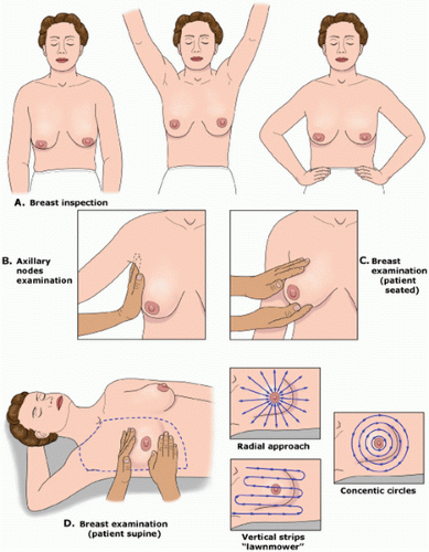
FIGURE 16.1 Clinical breast examination. A: The breast exam is started with the patient in a seated position with her arms relaxed. Breast inspection is aided by patient positioning. The patient is asked to raise her arms over her head so the lower part of the breasts can be inspected for asymmetry, skin changes, and nipple inversion or retraction. The patient then puts her hands on her hips and presses in to contract the pectoral muscles so that any other areas of retraction can be visualized. B: The regional lymph node exam is completed while the patient is still in the sitting position and includes the cervical, supraclavicular, infraclavicular, and axillary nodal basins. C: A bimanual examination of the breasts can be performed while the patient is still in the sitting position. This is especially useful for women with large pendulous breasts. D: The breast exam is completed with the patient in a supine position with the ipsilateral arm raised above her head. The area examined should extend from the clavicle superiorly to the rib cage inferiorly and from the sternum medially to the midaxillary line laterally. A systematic approach ensures that the entire breast is examined. This can be accomplished with either concentric circles, a radical approach, or vertical strips, referred to as the “lawnmower” method. (From Sabel MS. Breast masses and other common breast problems. UpToDate Web site. http://www.uptodate.com/contents/breast-masses-and-other-common-breast-problems. Accessed December 2, 2013.)


The nipple areola complex should be examined for changes in the skin, symmetry, and retraction. Newonset nipple retraction should be considered suspicious for malignancy. Ulceration or eczematoid lesions of the nipple areola complex can be a sign of Paget disease.

The patient should raise her arms over her head and then place her hands against her hips, resulting in contraction of the pectoralis muscles, which can demonstrate subtle nipple retraction and dimpling of the breast skin. With the patient still in the upright position, the regional lymph node basins, including cervical, supraclavicular, and axillary lymph nodes should be examined. Palpable lymph nodes should be characterized by their size, whether they are firm, mobile or fixed, or tender to palpation. Each breast should then be palpated by placing one hand under the breast supporting it, while using the flat portion of the other hand to examine the entire breast.

The patient is then placed in the supine position, and the ipsilateral arm is raised above the head. For patients with large pendulous breasts, a towel or pillow can be placed beneath the ipsilateral shoulder to elevate the breast that is being examined. The breast should be examined either in a concentric circle or in a radial pattern, and palpation should extend from the clavicle superiorly, laterally to the latissimus dorsi muscle (posterior axillary line), inferiorly to the costal margin, and medially to the sternum. This is performed with one hand placed on the breast to stabilize the breast, while the flat surface of the other hand is used to examine the breast. Palpation of the breast should include temperature differences of the surface of the skin, edema, focal or generalized tenderness, the presence of a dominant mass, and nipple discharge. Diffuse nodularity predominantly in the upper outer quadrants of the breast is a normal finding especially in premenopausal women. Comparing physical findings of both breasts is helpful in further differentiating nodularity or other areas of palpable concern. If there is an area of nodular breast tissue in a premenopausal woman that is nonspecific, the patient may be asked to return at a different time in her menstrual cycle for a physical examination. A dominant breast mass should be different from the surrounding breast tissue on examination. If a breast mass is noted, the size (in centimeters), location, and characteristics including tenderness, mobility, and firmness should be documented. It is helpful to record the location of any abnormality on the breast by documenting its position and the distance in centimeters from the areola because this will help on subsequent follow-up examinations by either the initial physician or other examiners. Diagrammatic depictions are quite useful, and many electronic medical records do have mechanisms for the creation of such figures. In addition, if a patient reports a palpable mass that is not found on physical examination, it is important to have the patient indicate the area of the palpable abnormality noted on breast self-examination. Physical examination alone will not distinguish between benign and cancerous lesions so any abnormal findings on examination should be further evaluated using imaging, for example, ultrasound or mammography, and if necessary, biopsy.


BREAST IMAGING


Mammography

Mammography uses low-dose radiation (0.3 rad/study) and is an effective screening tool for early detection of breast cancer. It is usually the first diagnostic test ordered in women older than the age of 30 years with a new breast complaint. Digital mammography is now widely used for both screening and diagnostic studies. In contrast to film mammography where the energy from the image receptors are captured on film, digital imaging uses a digital detector that is between the breast and the compression plate to convert the x-ray images electronically and project the images onto a monitor, allowing radiologists to manipulate and enhance the images seen.

In a study by Kerlikowske et al.,29 over 329,261 women between the age of 40 and 79 years underwent routine screening mammography. Two-thirds of the patients (638,252) had film mammography, and the remaining one-third of patients (231,034) had digital screening. Although the overall cancer detection rates were similar for digital and film mammography, the sensitivity and specificity of digital mammography was higher than film screening mammography in pre- and perimenopausal women (87.1 % versus 81.7%, respectively), women with extremely dense breasts (83.6% versus 68.1%, respectively), and women with estrogen-negative breast cancers (78.5% versus 65.8%, respectively).

Screening mammography is used to detect breast cancer in asymptomatic women, whereas diagnostic mammography is used to evaluate patients who present with clinical findings or an abnormal finding on a screening mammogram. Even if a woman has had a recent negative screening mammogram, if there is a focal complaint or change noted on exam, a diagnostic mammogram should be obtained. For screening mammography, two views of the breast are obtained: the craniocaudal (CC) and the mediolateral oblique (MLO). The MLO view is the most effective view because it includes the whole breast including the upper outer quadrant and the axillary tail. The CC view, which is performed using greater compression of the breast, provides better visualization of the medial aspect of the breast with better image detail. In addition to the MLO and the CC view, diagnostic mammograms use a 90-degree lateral view to
triangulate the exact location of the abnormality and spot compression views over the area of interest which allow for greater compression, displacing the overlying breast tissue that could obscure a lesion.

The American College of Radiology Breast Imaging Reporting and Data System (BI-RADS) uses standardized terminology to facilitate uniformity in mammography reports and includes an overall assessment of the probability of malignancy based on radiologic findings.30 There are six categories, each associated with a specific recommendation for management (Table 16.3).

Masses and calcifications are the most common abnormalities identified on mammography. In the final reporting of these findings, radiologists should indicate the likelihood of these findings being suspicious for malignancy. These findings should be detected on both the CC and the MLO view. Irregular or lobular-shaped lesions with indistinct or obscure margins are more likely to be associated with malignancy, and round, circumscribed lesions with well-defined margins are more likely to be benign findings.

Other findings on mammography that may be associated with a mass include nipple retraction, skin retraction, and skin thickening. Focal skin thickening can be associated with benign diseases such as mastitis or malignancy including inflammatory breast cancer. Architectural distortion is seen as an area of spiculation or retraction with distortion of normal landmarks, and unless this finding is associated with a prior biopsy site, a tissue diagnosis may be required to rule out malignancy.

Calcifications in the breast as small as 50 µm can be visible on mammography. Diffuse, scattered, coarse, large, and round calcifications are almost always benign. Pleomorphic, heterogeneous, fine, linear, and branching calcifications have a higher probability of being malignant.31, 32, 33 Microcalcifications associated with a mass are highly suggestive of a malignant lesion.34,35


TABLE 16.3 The American College of Radiology Breast Imaging Reporting and Data System


Category


Assessment


Follow-Up Recommendations


1


Negative


Routine annual screening mammography (for women over age 40)


2


Benign Finding(s)


Routine annual screening mammography (for women over age 40)


3


Probably Benign Finding — Initial Short-Interval Follow-Up Suggested


Initial short-term follow-up (usually 6-month) examination


4


Suspicious Abnormality — Biopsy Should Be Considered


Usually requires biopsy


5


Highly Suggestive of Malignancy — Appropriate Action Should Be Taken


Requires biopsy or surgical treatment


6


Known Biopsy-Proven Malignancy — Appropriate Action Should Be Taken


Category reserved for lesions identified on imaging study with biopsy proof of malignancy prior to definitive therapy


Mammography should be performed prior to any planned biopsy of a palpable mass in order to better define the palpable abnormality, and to evaluate the ipsilateral and contralateral breast for additional lesions. In addition, biopsies should be performed if there is a palpable mass, despite the absence of findings on mammography. Mammography has been reported to have an overall sensitivity of 50 to 90%.36, 37, 38 Young women who have more fibroglandular tissue or older women taking hormone replacement therapy who have extremely dense breasts will have lower sensitivity of mammography.39, 40, 41 Murphy et al.42 reported their experience of 1365 women with symptomatic breast cancer over a 10-year study period. They reported a false-negative rate of 10% in their study population, and these patients were more likely to be younger, have smaller tumors, and have tumors located outside the upper outer quadrant of the breast. A normal mammogram does not obviate the need for further evaluation of a suspicious mass. Mammography misses 10 to 20% of clinically palpable breast cancers.43 If a suspicious mass is found on examination, a negative mammogram does not represent a stopping point in the workup and evaluation.


Ultrasonography

Breast ultrasonography plays an important role in the evaluation of patients with breast diseases. Focused high frequency transducers provide reliable information using noninvasive techniques.44 It is an important adjunct to other imaging modalities such as mammography and magnetic resonance imaging (MRI). The benefit of routine screening ultrasonography has not been established; however, its role as a screening modality adjunct in women with dense breasts has been studied.45,46 In a clinical trial by the American College of Radiology Imaging Network, 2809 high-risk women older than the age of 25 years with extremely dense breasts had screening ultrasounds in conjunction with mammography.47 This study demonstrated that the addition of ultrasound was significantly associated with a higher likelihood of diagnosing breast cancer (11.8 per 1000 women) when compared to mammography alone (7.6 per 1000); however, the positive predictive value was low. Only 20 (8.6%) of the 233 women who were recommended to undergo a biopsy were diagnosed with breast cancer with over 90% of patients having benign findings. Other studies confer similar false-positive rates with the use of ultrasound and mammography in women with radiographically dense breasts.48, 49, 50 The improved diagnostic yield in this population of women should be weighed against the anxiety women face from high false-positive rates and unnecessary biopsies.


Ultrasound does have reliable criteria that can help distinguish a benign cyst from a malignant solid mass.51,52 If a lesion seen on ultrasound is a simple cyst, then no further imaging or intervention is needed. Lesions that are solid and irregular and have ill-defined margins are more likely to be malignant, and a core needle biopsy (CNB) should be performed.

The American College of Radiology (ACR) criteria for breast ultrasonography in the management of women with breast disease includes evaluation of a palpable mass or other breast-related symptoms, evaluation of lesions seen on other imaging modalities such as mammography and MRI, imaging of women younger than age 30 years or women who are lactating or pregnant, implant leaks, obscure mammographic findings in women with dense breasts, and for guidance for interventional procedures.


Magnetic Resonance Imaging

MRI provides both high spatial and temporal resolution to provide three-dimensional imaging that differentiates benign versus malignant lesions. Breast MRI has the advantage of being highly sensitive, with the ability to detect cancers that are not visualized on mammography or ultrasonography without the use of ionizing radiation.53, 54, 55 The main disadvantages include its low specificity, high cost, and prolonged imaging time (45 minutes). Although gadolinium contrast enhancement kinetics is used to differentiate benign from malignant enhancement, benign lesions may also demonstrate contrast enhancement similar to enhancement of malignant lesions, leading to unnecessary biopsies.56, 57, 58 A baseline blood urea nitrogen (BUN) and creatinine should be checked prior to the use of gadolinium dye because it may cause serious reactions in patients with underlying renal disease (e.g., nephrogenic systemic fibrosis and worsening renal failure).

Several large studies have demonstrated that patients most likely to benefit from breast MRI are women with breast cancer and women who are at high risk of developing breast cancer. Lesions seen with MRI may not be apparent on other imaging modalities.59 In women newly diagnosed with breast cancer, MRI identifies additional ipsilateral disease in patients with presumed unifocal disease. This is most often observed in premenopausal women, women with dense breasts on mammography, patients with larger lesions (greater than 5 cm), those with a family history of breast cancer, and women diagnosed with invasive lobular carcinoma.60, 61, 62 The controversy remains whether all women with a new diagnosis of breast cancer should undergo preoperative MRI given the cost. In addition, several retrospective nonrandomized trials have shown no significant differences in overall survival or disease-free survival in women with breast cancer who underwent preoperative MRI when compared to a group that did not have a staging MRI prior to breast-conserving surgery.63, 64, 65

MRI can be a useful clinical tool in the management of occult primary breast cancer patients who have malignant axillary adenopathy with a normal breast exam, mammogram, and ultrasound.66,67 In addition, MRI is the most accurate imaging tool available to monitor the response to chemotherapy in patients undergoing neoadjuvant chemotherapy.68,69 The role for MRI as routine surveillance in women with a personal history of breast cancer remains unknown; however, MRI can be useful to differentiate surgical changes versus recurrent disease in women who have undergone breast conserving surgery and have indeterminate mammographic findings at the lumpectomy site.

The American Cancer Society has recommended guidelines for MRI in addition to screening mammography in high-risk women. These categories include women who have a 20 to 25% or greater lifetime risk of developing breast cancer as determined by risk assessment tools, women with a known genetic mutation, women who have not been tested but have a known genetic mutation in the family, and women who have previously received chest wall radiation.70


Thermography

Breast thermography has been proposed as a method of distinguishing benign and malignant lesions and as a potential screening tool in high-risk women. Temperature maps of the breast are created using various devices that measure the heat patterns and number of vessels between both breasts, the temperature of both the skin surface and the vasculature, and the dynamic changes in breast temperature over time. Variations in the production of heat by the breast have been proposed to indicate breast disease.71,72 There are multiple small trials evaluating the metabolic activity of malignant and benign breast lesions; however, the potential benefits as a screening tool in the detection of cancer is unknown and at this time should not be performed. In addition, the National Institutes of Health (NIH) has reported as a consensus statement that there is no data on the efficacy of thermography, and it should not be used as a screening technique for women.


DIAGNOSTIC TECHNIQUES

All suspicious lesions on clinical breast exam or nonpalpable abnormalities detected on imaging requires appropriate diagnostic evaluation using needle biopsy, either fine needle aspiration, or preferably CNB, or less commonly, surgical excision with preoperative needle localization. Excision should be reserved only for lesions not amenable to image-guided core biopsy. Figure 16.2 presents an algorithm for the management of a breast mass.


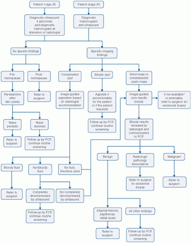
FIGURE 16.2 Algorithm for the management of a breast mass. PCP, primary care provider.


Fine Needle Aspiration

Fine needle aspiration (FNA) requires the use of a handheld syringe to percutaneously aspirate cells for cytologic evaluation. In multiple large studies, the sensitivity has varied from 65 to 91% and specificity from 46 to 100%.73, 74, 75 Although FNA is better tolerated than CNB and enables a pathologist to immediately ascertain the adequacy of the sample, it requires a skilled cytopathologist to interpret the results. In addition, FNA is subject to sampling error because lesions are aspirated with a small 20- or 22-gauge needle, and even if adequate sampling is obtained, cytology cannot differentiate in situ or invasive carcinoma. The diagnosis is described in five categories: (a) benign with no evidence of atypia or malignancy, (b) atypia/indeterminate (nondiagnostic cellular material), (c) suspicious for malignancy, (d) malignant, (e) material is unsatisfactory for evaluation. The combination of cytology, clinical breast exam, and imaging must be correlated to achieve triple test concordance.76 A negative FNA in the presence of findings of a suspicious mass on clinical breast exam or imaging should prompt further investigation. For patients with a diagnosis of “suspicious” or “probably malignant,” a CNB or excision should be performed before definitive surgical treatment to rule out a false-positive diagnosis.77 FNA has now largely been replaced by CNB.


Core Needle Biopsy

Over 1.4 million breast biopsy procedures are performed each year in the United States; however, a significant portion of these are performed for benign disease.78 CNBs for palpable lesions or the use of image guidance for nonpalpable lesions is the diagnostic procedure of choice and has virtually eliminated open surgical biopsy and FNA. Image guidance can be performed using ultrasound or mammography. CNB is minimally invasive and cost-effective compared to surgical excision for diagnostic purposes. Stereotactic biopsies are performed for suspicious calcifications or density seen only on mammography and not ultrasonography. Core biopsies are associated with more discomfort than FNA but provide better tissue sampling and the ability to classify subtypes of cancer. Most procedures are being performed with either 10-, 11-, or 14-gauge devices. Vacuum-assisted devices can also be used to provide large cores enabling a more extensive sample.79, 80, 81

One of the challenges of CNB is the accuracy of distinguishing certain histopathologic entities. Some studies have demonstrated an inability to distinguish atypical hyperplasia from low-grade ductal carcinoma in situ (DCIS). The findings of atypia or a papillary lesion such as a papilloma warrants excision with upstaging to carcinoma seen in 4 to 20% of patients.82, 83, 84 In addition, other investigators have demonstrated pathologic upstaging of benign papillary lesions on CNB to atypical ductal hyperplasia or carcinoma on final surgical excision.85, 86, 87


Incisional/Excisional Biopsy

Incisional biopsy refers to surgical excision of a portion of the lesion, whereas excisional biopsy results in surgical removal of the entire lesion. Incisional biopsies used to be the procedure of choice for diagnosing large palpable lesions; however, CNB has virtually eliminated the need for this procedure.

Excisional biopsy is only used when image-guided CNB is technically not feasible, if CNB results are discordant with clinical findings, if FNA or CNB is nondiagnostic, or if a CNB demonstrates atypical ductal/lobular hyperplasia or a papilloma.


Breast Ductoscopy/Cytology

Ductal lavage was developed as a method of collecting ductal epithelial cells from samples obtained through cannulating a duct with a microcatheter, inserting small amounts of saline, and through gentle suction, nipple aspirate is performed for cytologic evaluation. The procedure was designed to detect abnormal epithelial cells in high-risk women who had normal clinical breast exams and imaging. Ductal lavage has the ability to detect abnormal intraductal epithelial cells; however, the information gained from these results in women who have no findings on breast exam or imaging is less clear. It is not being used as a diagnostic clinical tool at the present time but is being performed in research protocols.


BENIGN BREAST CONDITIONS

Benign breast lesions are the most common presenting breast disorder seen by clinicians. Patients with benign lesions are often managed to exclude breast cancer rather than a necessity to understand and manage each of these entities.


Fibrocystic Change

Fibrocystic changes, also known as fibrocystic disease, is the most common lesion in the breast and accounts for nearly 20 to 25% of all breast lesions. Pathologists describe this entity as fibrosis with apocrine cysts and epithelial hyperplasia. Histologic findings are now classified into one of three categories that are strictly defined and correlate with the risk of developing breast cancer: non-proliferative lesions, proliferative lesions without atypia, and atypical hyperplasias. Cysts can range in size from 1 mm to several centimeters and arise as a result of individual acini or ducts that dilate and unfold and enlarge as a cyst. Haagensen89 first described the coalescing of clusters of cysts to form a gross cyst that was palpable. Estrogen seems to play a role in producing symptoms because this entity most commonly occurs between the
age of 35 and 50 years and is extremely unusual in postmenopausal women not on estrogen replacement.


Clinical Findings

Fibrocystic changes usually produce a tender mass that is smooth, mobile, and compressible. Pain is the most common symptom and is usually associated with the menstrual cycle when cysts are known to enlarge from hormonal stimulation. In addition, fluctuations in size are often noted at various times in the menstrual cycle. Cysts can occur in multiple areas within the same breast and bilaterally with cyclical breast pain. Quite commonly, cyclical breast pain is focal without the detection of cysts on imaging or physical examination, and no cause can be identified.


Diagnostic Tests

Clinically, women present with a mass that produces significant anxiety and can be hard to distinguish from cancer. Mammography may be normal in these patients, and there are no specific radiographic criteria that help to distinguish this entity. Ultrasound is the most useful test in differentiating a cyst from a solid mass (Fig. 16.3A & B). Findings on ultrasound characteristic of a simple cyst include a round lesion with smooth borders, posterior acoustic enhancement, and absence of internal echoes. If ultrasound findings are not classic for a simple cyst and demonstrate internal septations within the cyst, the cyst is classified as a complex cyst, and FNA or CNB should be performed for diagnosis.

Most patients need reassurance and do not require further treatment after ultrasound confirmation of a simple cyst or for cyclical pain with no findings. Aspiration of a simple cyst is only performed if a patient is symptomatic. Aspiration is usually performed with an 18- or 20-gauge needle, and if nonbloody fluid is aspirated, the fluid is not sent for cytology. If aspiration results in complete resolution of the cyst, patients can be clinically followed. If under ultrasound guidance the cyst continues to recur despite repeated aspirations, a mass is noted after aspiration of the cyst, or if bloody fluid is aspirated, needle biopsy or excision is recommended due to the risk of atypia or malignancy.

Dietary modulation and nutritional supplements have been investigated in providing symptomatic relief. The role of caffeine and other caffeine-containing products including tea and chocolate in accentuating pain remains controversial.90,91 Some patients do report pain relief after discontinuing caffeine-containing products. Similarly, observational studies have shown vitamin E (600 IU daily) or B6 (200 to 800 mg/day) may be helpful in reducing symptoms.92,93


Fibrocystic Changes and Cancer Risk

Fibrocystic change that demonstrates apocrine changes with mild epithelial hyperplasia of usual type does not increase future risk of breast cancer; however, moderate or florid hyperplasia with or without atypia on biopsy is associated with an increased risk of breast cancer.94,95 Women with proliferative changes and no atypical hyperplasia have a two-fold risk of developing breast cancer. Patients with biopsies that demonstrate proliferative changes with atypical hyperplasia have a four- to five-fold risk of developing breast cancer in either breast. A family history of breast cancer in a patient with cysts and proliferative changes increases the risk three-fold. Women with proliferative changes and family history should be followed with clinical breast exams and mammography annually.


FIGURE 16.3 A: Ultrasound demonstrating a simple cyst. B: Ultrasound demonstrating a solid mass.


Mastalgia

Breast pain remains one of the most common reason patients seek medical care due to concerns about risk of breast cancer. It is a physiologic symptom that frequently occurs in the luteal phase of the menstrual cycle. Localized breast pain may be the only presenting symptom in up to 15% of women with newly diagnosed
breast cancer.96 Research has focused on abnormal levels of estradiol, progesterone, and prolactin as possible causes of mastalgia; however, no consistent findings have been observed.97, 98, 99 Inflammatory cytokines, interleukin-6, and tumor necrosis-α have also been implicated; however, no differences in expression have been seen in breast tissue from women who have no pain when compared to breast tissue from women who have mastalgia.100

Only gold members can continue reading. Log In or Register to continue

Stay updated, free articles. Join our Telegram channel

Jun 25, 2016 | Posted by in GYNECOLOGY | Comments Off on Breast Disorders

Full access? Get Clinical Tree

Get Clinical Tree app for offline access