Blood Disorders

Chapter 97 Blood Disorders


97.1 Anemia in the Newborn Infant


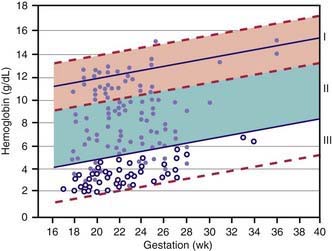
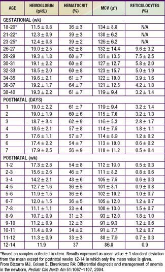
Hemoglobin increases with advancing gestational age: at term, cord blood hemoglobin is 16.8 g/dL (14-20 g/dL); hemoglobin levels in very low birthweight (VLBW) infants are 1-2 g/dL below those in term infants (Fig. 97-1). A hemoglobin value less than the normal range of hemoglobin for birthweight and postnatal age is defined as anemia (Table 97-1). A “physiologic” decrease in hemoglobin content is noticed at 8-12 wk in term infants (hemoglobin, 11 g/dL) and at about 6 wk in premature infants (7-10 g/dL).


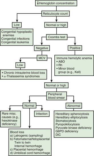
Infants born by cesarean section may have a lower hematocrit (Hct) than those born vaginally. Anemia at birth is manifested as pallor, heart failure, or shock (Fig. 97-2). It may be due to acute or chronic fetal blood loss, hemolysis, or underproduction of erythrocytes. Specific causes include hemolytic disease of the newborn, tearing or cutting of the umbilical cord during delivery, abnormal cord insertion, communicating placental vessels, placenta previa or abruptio, nuchal cord, incision into the placenta, internal hemorrhage (liver, spleen, intracranial), α-thalassemia, congenital parvovirus infection or other hypoplastic anemias, and twin-twin transfusion in monozygotic twins with arteriovenous placental connections (Chapter 92).


Transplacental hemorrhage with bleeding from the fetal into the maternal circulation has been reported in 5-15% of pregnancies, but, unless severe, it is not usually sufficient to cause clinically apparent anemia at birth. The cause of transplacental hemorrhage is not clear, but its occurrence has been proven by demonstration of significant amounts of fetal hemoglobin and red blood cells (RBCs) in maternal blood on the day of delivery by the Kleihauer-Betke test or by flow cytometry methods to detect fetal cells in maternal blood. If the infant has severe anemia with heart failure, emergency exchange transfusion to restore Hct and oxygen-carrying capacity may be needed.


Acute blood loss usually results in severe distress at birth, initially with a normal hemoglobin level, no hepatosplenomegaly, and early onset of shock. In contrast, chronic blood loss in utero produces marked pallor, less distress, a low hemoglobin level with microcytic indices, and, if severe, heart failure.


Anemia appearing in the first few days after birth is also most frequently a result of hemolytic disease of the newborn. Other causes are hemorrhagic disease of the newborn, bleeding from an improperly tied or clamped umbilical cord, large cephalohematoma, intracranial hemorrhage, and subcapsular bleeding from rupture of the liver, spleen, adrenals, or kidneys. Rapid decreases in hemoglobin or Hct values during the first few days of life may be the initial clue to these conditions.


Later in the neonatal period, delayed anemia may develop as a result of hemolytic disease of the newborn, with or without exchange transfusion or phototherapy. Congenital hemolytic anemia (spherocytosis) occasionally appears during the 1st mo of life, and hereditary nonspherocytic hemolytic anemia has been described during the neonatal period secondary to deficiency of glucose-6-phosphate dehydrogenase (G6PD) and pyruvate kinase. Bleeding from hemangiomas of the upper gastrointestinal tract or from ulcers caused by aberrant gastric mucosa in a Meckel diverticulum or duplication is a rare source of anemia in newborns. Repeated blood sampling of infants requiring frequent monitoring of blood gas and chemistry parameters is a common cause of anemia among hospitalized infants. Deficiency of minerals such as copper may cause anemia in infants maintained on total parenteral nutrition.


Anemia of prematurity occurs in LBW infants 1-3 mo after birth, is associated with hemoglobin levels <7-10 g/dL, and is clinically manifested as pallor, poor weight gain, decreased activity, tachypnea, tachycardia, and feeding problems. Repeated phlebotomy for blood tests, shortened RBC survival, rapid growth, and the physiologic effects of the transition from fetal (low PaO2 and hemoglobin saturation) to neonatal life (high PaO2 and hemoglobin saturation) contribute to anemia of prematurity. The oxygen available to neonatal tissue is lower than that in adults, but a neonate’s erythropoietin response is attenuated for the degree of anemia, and as a result, hemoglobin and reticulocyte levels are low. In VLBW infants, delayed clamping of the umbilical cord with the infant held below the level of the placenta may enhance placental-infant transfusion and reduce postnatal transfusion needs. This maneuver should not delay any needed resuscitation and may lead to hyperviscosity.


Delayed cord clamping (≈1-2 min or after cessation of cord pulsation) may be beneficial in otherwise well newborns in preventing anemia in full-term infants, with effects extending beyond the neonatal period. The benefits of delayed cord clamping persist for 2-6 mo as improved hematocrit, iron status as measured by ferritin concentration and stored iron, and a clinically important reduction in the risk of anemia in infancy. Late clamping may result in delivery of an extra 20-40 mL of blood and 30-35 mg of iron to the newborn. Polycythemia is a risk with delayed clamping but is often asymptomatic.


Treatment of neonatal anemia by blood transfusion depends on the severity of symptoms, the hemoglobin level, and the presence of co-morbid diseases (bronchopulmonary dysplasia, cyanotic congenital heart disease, respiratory distress syndrome) that interfere with oxygen delivery. The need for treatment with blood should be balanced against the risks of transfusion, including hemolytic transfusion reactions, exposure to blood product preservatives and other potential toxins, volume overload, possible increased risk of retinopathy of prematurity and necrotizing enterocolitis, graft-versus-host (GVH) reaction, and transfusion-acquired infection (cytomegalovirus [CMV], HIV, parvovirus, hepatitis B and C) (Chapter 468). The risk of CMV infection can be almost eliminated by the use of leukoreduced blood. In the infant <1,500 g, CMV antibody-negative leukoreduced blood should be used. The risk of acquiring HIV and hepatitis B and C viruses is reduced but not eliminated by antibody screening of donated blood. Blood banking techniques that limit multiple donor exposure should be encouraged.


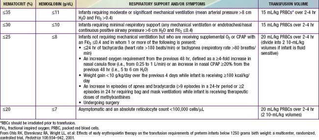
Although transfusion guidelines for preterm infants have been proposed (Table 97-2), they have not been subjected to rigorous clinical study. Nonetheless, these guidelines have led to a decline in the number of unnecessary transfusions. The use of restrictive vs more liberal transfusion guidelines has been examined in two randomized trials, one conducted at University of Iowa and a second multicentric trial known as the PINT (Premature Infants in Need of Transfusion) study. The restrictive guidelines in the two groups were generally similar. In the Iowa trial, the transfusion thresholds in the liberal- and restrictive-transfusion groups were <46% and <34%, respectively, in tracheally intubated infants receiving assisted ventilation; <38% and <28%, respectively, in infants receiving nasal continuous positive airway pressure or supplemental oxygen; and <30% and <22%, respectively, in infants breathing room air. The transfusion thresholds for the liberal groups were higher in the Iowa trial than in the PINT study. In both trials, the use of restrictive thresholds resulted in fewer transfusions and also increased the number of infants who received no transfusions at all. However, in the Iowa trial (but not in the PINT study), restrictive transfusion thresholds were associated with increases in major cranial ultrasonographic abnormalities and in the frequency of apneic spells. Although these findings need further evaluation in clinical studies, the issue of finding an appropriate transfusion threshold in premature infants remains unresolved.


Asymptomatic full-term infants with a hemoglobin level of 10 g/dL may be monitored, whereas symptomatic neonates born after abruptio placentae or with severe hemolytic disease of the newborn need immediate transfusion. Preterm infants who have repeated episodes of apnea and bradycardia despite theophylline therapy and a hemoglobin level ≤8 g/dL may benefit from RBC transfusion. In addition, infants with respiratory distress syndrome or severe bronchopulmonary dysplasia may need hemoglobin levels of 12-14 g/dL to improve oxygen delivery. No transfusion is needed to replace blood removed for testing or for mild asymptomatic anemia. Asymptomatic neonates with reticulocytopenia and hemoglobin levels ≤7 g/dL may require transfusion; if a transfusion is not provided, close observation is essential. Packed RBC transfusion (10-20 mL/kg) is given at a rate of 2-3 mL/kg/hr to raise the hemoglobin concentration; 2 mL/kg raises the hemoglobin level 0.5-1 g/dL. Hemorrhage should be treated with whole blood if available; alternatively, fluid resuscitation is initiated, followed by packed RBC transfusion.


Recombinant human erythropoietin (r-HuEPO) may be considered in the treatment of chronic or anticipated anemia in an attempt to decrease or eliminate transfusions when families, for religious reasons, request all possible measures to avoid transfusions. Therapy with r-HuEPO must be supplemented with oral iron. Doses and regimens vary. In anemia of prematurity, r-HuEPO does not provide a major reduction in transfusion requirements or the number of donors; therefore, routine use of erythropoietin in VLBW infants is not recommended. Early initiation of r-HuEPO therapy may produce a small reduction in the total transfusion volume per infant. There were concerns about an increased risk of severe retinopathy of prematurity in the r-HuEPO group. The effects of late initiation of r-HuEPO (≥8 days) have also been associated with small reductions in the total blood volume transfused per infant and the number of transfusions per infant.


Bibliography


Aher S, Ohlsson A: Late erythropoietin for preventing red blood cell transfusion in preterm and/or low birth weight infants, Cochrane Database Syst Rev (3):CD004868, 2006.


Anderson C. Critical haemoglobin thresholds in premature infants. Arch Dis Child Fetal Neonatal Ed. 2001;84:F146-F148.


Bell EF, Strauss RG, Widness JA, et al. Randomized trial of liberal versus restrictive guidelines for red blood cell transfusion in preterm infants. Pediatrics. 2005;115:1685-1691.


Bizzarro MJ, Colson E, Ehrenkranz RA. Differential diagnosis and management of anemia in the newborn. Pediatr Clin North Am. 2004;51:1087-1107.


Christensen RD, Henry E. Hereditary spherocytosis on neonates with hyperbilirubinemia. Pediatrics. 2010;125:120-125.


Ferguson D, Hébert PC, Lee SK, et al. Clinical outcomes following institution of universal leukoreduction of blood transfusions for premature infants. JAMA. 2003;289:1950-1956.


Hébert PC, Fergusson D, Blajchman MA, et al. Clinical outcomes following institution of the Canadian universal leukoreduction program for red blood cell transfusions. JAMA. 2003;289:1941-1949.


Hutton EK, Hassan ES. Late vs early clamping of the umbilical cord in full-term neonates. JAMA. 2007;297:1241-1252.


Kirpalani H, Whyte RK, Andersen C, et al. The Premature Infants in Need of Transfusion (PINT) study: a randomized, controlled trial of a restrictive (low) versus liberal (high) transfusion threshold for extremely low birth weight infants. J Pediatr. 2006;149:301-307.


Nicaise C, Gire C, Casha P, et al. Erythropoietin as treatment for late hyporegenerative anemia in neonates with Rh hemolytic disease after in utero exchange transfusion. Fetal Diagn Ther. 2002;17:22-24.


Ohlsson A, Aher SM: Early erythropoietin for preventing red blood cell transfusion in preterm and/or low birth weight infants, Cochrane Database Syst Rev (3):CD004863, 2006.


Rabe H, Reynolds G, Diaz-Rossello J: Early versus delayed umbilical cord clamping in preterm infants, Cochrane Database Syst Rev (4):CD003248, 2004.


97.2 Hemolytic Disease of the Newborn (Erythroblastosis Fetalis)


Erythroblastosis fetalis is caused by the transplacental passage of maternal antibody active against paternal RBC antigens of the infant and is characterized by an increased rate of RBC destruction. It is an important cause of anemia and jaundice in newborn infants despite the development of a method of preventing maternal isoimmunization by Rh antigens. Although more than 60 different RBC antigens are capable of eliciting an antibody response, significant disease is associated primarily with the D antigen of the Rh group and with incompatibility of ABO factors. Rarely, hemolytic disease may be caused by C or E antigens or by other RBC antigens, such as CW, CX, DU, K (Kell), M, Duffy, S, P, MNS, Xg, Lutheran, Diego, and Kidd. Anti-Lewis antibodies do not cause disease.


Hemolytic Disease of the Newborn Caused by Rh Incompatibility


The Rh antigenic determinants are genetically transmitted from each parent, determine the Rh type, and direct the production of a number of blood group factors (C, c, D, d, E, and e). Each factor can elicit a specific antibody response under suitable conditions; 90% are due to D antigen and the remainder to C or E antigens.


Pathogenesis


Isoimmune hemolytic disease from D antigen is approximately three times more frequent among white persons than among black persons. When Rh-positive blood is infused into an Rh-negative woman through error or when small quantities (usually > 1 mL) of Rh-positive fetal blood containing D antigen inherited from an Rh-positive father enter the maternal circulation during pregnancy, with spontaneous or induced abortion, or at delivery, antibody formation against D antigen may be induced in the unsensitized Rh-negative recipient mother. Once sensitization has taken place, considerably smaller doses of antigen can stimulate an increase in antibody titer. Initially, a rise in immunoglobulin (Ig) M antibody occurs, which is later replaced by IgG antibody; the latter readily crosses the placenta to cause hemolytic manifestations.


Hemolytic disease rarely occurs during a first pregnancy because transfusion of Rh-positive fetal blood into an Rh-negative mother occurs near the time of delivery, too late for the mother to become sensitized and transmit antibody to her infant before delivery. The facts that 55% of Rh-positive fathers are heterozygous (D/d) and may have Rh-negative offspring and that fetal-to-maternal transfusion occurs in only 50% of pregnancies reduce the chance of sensitization, as does small family size, in which the opportunities for its reoccurrence are reduced. The disparity between the numbers of incompatible vs alloimmunized maternal-fetal pairs can also be due to a threshold effect of fetomaternal transfusions (a certain amount of the immunizing blood cell antigen is required to activate the maternal immune system), the type of antibody response (IgG antibodies are more efficiently transferred across the placenta to the fetus), differential immunogenicity of blood group antigens, and differences in maternal immune response, presumably related to differences in the efficiency of antigen presentation by various major histocompatibility loci. Thus, the overall incidence of isoimmunization of Rh-negative mothers at risk is low, with antibody to antigen D detected in >10% of those studied, even after five or more pregnancies; only about 5% ever have babies with hemolytic disease.


When the mother and fetus are also incompatible with respect to group A or B, the mother is partially protected against sensitization by the rapid removal of Rh-positive cells from her circulation by her preexisting anti-A or anti-B antibodies, which are IgM antibodies and do not cross the placenta. Once a mother has been sensitized, her infant is likely to have hemolytic disease. The severity of Rh illness worsens with successive pregnancies. The possibility that the first affected infant after sensitization may represent the end of the mother’s childbearing potential for Rh-positive infants argues urgently for the prevention of sensitization. The injection of anti-D gamma globulin (RhoGAM) into the mother immediately after the delivery of each Rh-positive infant has been a successful strategy to reduce Rh hemolytic disease (see later).


Clinical Manifestations


A wide spectrum of hemolytic disease occurs in affected infants born to sensitized mothers, depending on the nature of the individual immune response. The severity of the disease may range from only laboratory evidence of mild hemolysis (15% of cases) to severe anemia with compensatory hyperplasia of erythropoietic tissue leading to massive enlargement of the liver and spleen. When the compensatory capacity of the hematopoietic system is exceeded, profound anemia occurs and results in pallor, signs of cardiac decompensation (cardiomegaly, respiratory distress), massive anasarca, and circulatory collapse. This clinical picture of excessive abnormal fluid in two or more fetal compartments (skin, pleura, pericardium, placenta, peritoneum, amniotic fluid), termed hydrops fetalis, frequently results in death in utero or shortly after birth. With the use of anti-D gamma globulin to prevent Rh sensitization, nonimmune (nonhemolytic) conditions have become frequent causes of hydrops (Table 97-3). The severity of hydrops is related to the level of anemia and the degree of reduction in serum albumin (oncotic pressure), which is due in part to hepatic dysfunction. Alternatively, heart failure may increase right heart pressure, with the subsequent development of edema and ascites. Failure to initiate spontaneous effective ventilation because of pulmonary edema or bilateral pleural effusions results in birth asphyxia; after successful resuscitation, severe respiratory distress may develop. Petechiae, purpura, and thrombocytopenia may also be present in severe cases as a result of decreased platelet production or the presence of concurrent disseminated intravascular coagulation.


Table 97-3 ETIOLOGY OF HYDROPS FETALIS*


Only gold members can continue reading. Log In or Register to continue

Stay updated, free articles. Join our Telegram channel

Jun 18, 2016 | Posted by in PEDIATRICS | Comments Off on Blood Disorders

Full access? Get Clinical Tree

Get Clinical Tree app for offline access
CATEGORY DISORDER(S)
Anemia Immune (Rh, Kell) hemolysis
α-Thalassemia
Red blood cell enzyme deficiencies (glucose-6-phosphate dehydrogenase)
Fetomaternal hemorrhage
Donor in twin-to-twin transfusion
Diamond-Blackfan syndrome
Cardiac dysrhythmias Supraventricular tachycardia