Benign Disorders of the Vulva and Vagina


Benign Disorders of the Vulva and Vagina


Colleen Kennedy Stockdale

Lori A. Boardman


INTRODUCTION

Vulvovaginal symptoms are common, often chronic, and can significantly interfere with sexual function and sense of well-being. Despite this, vulvovaginal symptoms are often underreported by women. Thus, when identified, vulvovaginal symptoms should be addressed by providers (including referral) to optimize care. In clinics specializing in the evaluation of women with vulvar complaints, the most common symptoms reported were vulvar pain, dyspareunia, and pruritus, with dermatologic conditions and vulvodynia (both generalized and localized forms) leading the diagnoses rendered.1

Understanding of the differences in epithelial and glandular structure, hormonal responsiveness, neural distribution, and immune responses is necessary when assessing vulvar complaints. For instance, the nonkeratinized vestibule is more permeable than keratinized skin, whereas the labia majora displays elevated hydration, occlusion, and frictional properties, all of which may increase irritant (chemical and mechanical) susceptibility.

Important vulvar landmarks include the labia majora, prominent hair-bearing folds of skin that represent the lateral boundaries of the vulva and meet to form the anterior boundary of the vulva at the mons pubis. Fusion of the labia majora at the fourchette determines the posterior boundary of the vulva. The non-hair-bearing labia minora lie medial to the labia majora and fuse anteriorly to form the prepuce and frenulum of the clitoris and thus mark the superior boundary of the vestibule (opening to the vagina). The vestibule is bound medially by the hymen and laterally by Hart’s line, a variably distinct line of demarcation evident at the base of the medial aspect of each labium minus. Hart’s line separates the nonkeratinized squamous epithelium of the vestibule from the keratinized epithelium of the more lateral labia minora and serves as an important landmark for several of the disorders discussed (Fig. 14.1).

When taking a history, it is important to ask about the onset, duration, location, and nature of vulvar symptoms (including the relationship of symptoms to the patient’s menstrual cycle) as well as any possible precipitating factors (including medications employed to reduce symptoms). In the evaluation of infectious causes of vulvovaginal complaints, microscopy, in conjunction with vaginal pH determination, helps guide the use of subsequent diagnostic tools, including vaginal fungal cultures, a variety of U.S. Food and Drug Administration (FDA)-approved point-of-care tests, culture or polymerase chain reaction (PCR) for confirmation of herpes simplex virus, and specific serologic tests.2,3 Examination of other skin sites such as the mouth, eyes, and scalp should also be done as nongenital-based disease may present with manifestations in the vulvar or vaginal areas. For autoimmune disorders and suspected cases of neoplasia, biopsy is invaluable.


Pruritus

In evaluating vulvar pruritus, it can be helpful to group women into those with acute versus chronic symptoms. Acute anogenital pruritus is most often infectious (as covered in Chapter 4: Vulvovaginitis and Chapter 6: Sexually Transmitted Infections and Pelvic Inflammatory Disease) with allergic and irritant contact dermatitis playing a role in some cases. Chronic pruritus, in general, has a history of gradual onset; examples of which include lichen simplex chronicus, lichen sclerosus, psoriasis, and various manifestations of human papillomavirus-related disease.


RECURRENT VULVOVAGINAL CANDIDIASIS

Candidiasis is the most common chief complaint by women presenting with vulvovaginal symptoms. A thorough history and evaluation are imperative for diagnosis. Most candidal infections are sporadic and, in more than 90% of cases, are caused by Candida albicans. Women with recurrent disease present a challenge both in terms of evaluation and treatment. Although testing for diabetes or immunosuppression is often suggested, such testing is rarely helpful. On the other hand, host factors, such as uncontrolled diabetes, immunosuppression, repeated exposure to antibacterials, and exogenous estrogen exposure may all be responsible for recurring episodes. Although non-albicans species should be suspected and culture is clearly warranted in this population, it should be remembered that repeated vulvovaginitis most commonly
occurs with persistent C. albicans, with non-albicans Candida species seen in approximately 10 to 20% of women with recurrent disease.


FIGURE 14.1 Normal anatomy. (From Beckmann CRB, Ling FW, Barzansky BM, et al. Obstetrics and Gynecology. 5th ed. Philadelphia: Lippincott Williams & Wilkins; 2006.)

A reliable diagnosis cannot be made based on symptoms or signs in the absence of corroborative laboratory data. In addition to direct microscopy, patients should have vaginal cultures obtained on at least two occasions, preferably while symptomatic. If two consecutively obtained cultures are negative for Candida species, the diagnosis of recurrent disease can be confidently excluded and other infectious (e.g., recurrent bacterial vaginosis, resistant trichomoniasis) and noninfectious (contact dermatitis, atrophic vaginitis, desquamative inflammatory vaginitis) etiologies should be considered.4

Recurrent infection with C. albicans can be treated with the same regimen used for severe disease (a 7- to 14-day course of a topical azole or two sequential doses of fluconazole), followed by 6 months of maintenance therapy with either oral medication (weekly fluconazole) or clotrimazole (weekly suppository).5 In a recent randomized controlled trial of suppressive therapy with fluconazole, women with recurrent disease on maintenance medication for 6 months, compared with those on placebo, were significantly more likely to remain disease-free at both 6 months (91% versus 36%) and at follow-up off medication 6 months later (43% versus 22%).5

Non-albicans infections (e.g., Candida glabrata, Candida krusei, Candida parapsilosis, Candida tropicalis) typically do not respond to treatment with fluconazole, often secondary to an intrinsic resistance to this medication. Recommended therapy, then, is a 7- to 14-day course of a nonfluconazole azole. If treatment fails, a 2-week course of nightly boric acid vaginal suppositories (600 mg in a 0 gel capsule) or nightly 4% flucytosine vaginal cream (requires a compounding pharmacy) can be used.6


VULVOVAGINAL EPITHELIAL DISORDERS


Human Papillomavirus-Related Disease

Genital human papillomavirus (HPV), the most common sexually transmitted viral infection, is associated with a number of vulvar and vaginal epithelial disorders, including genital warts, vulvar and vaginal intraepithelial neoplasia, and some carcinomas. Of the more than 120 HPV subtypes identified, more than 40 are specific to the anogenital tract, and of those, at least 15 are believed to be oncogenic. Commercially available probes for HPV DNA typing allow for identification of half of these anogenital subtypes, including 5 low-risk and 14 oncogenic subtypes. Although low-risk HPV types 6 and 11 are implicated in the development of genital warts and oncogenic HPV 16 commonly is found in classic (bowenoid) vulvar intraepithelial neoplasia (VIN), routine use of HPV testing for diagnosing vulvar HPV-related disease is not recommended at this time.

Estimates of the prevalence of HPV infection cannot be derived from visible manifestations of disease alone because the majority of infections are subclinical. Approximately 1% of women will develop condyloma, and few will develop dysplasia. Estimates suggest that HPV infection is associated with approximately 40% of vulvar cancers, compared to nearly 100% of cervical squamous cell cancers.7 In contrast to the typical keratinizing vulvar squamous carcinomas seen in older women (in which oncogenic HPV infection ranges from 2 to 23%), HPV-related basaloid and warty carcinomas (75 to 100% of which are associated with oncogenic HPV) tend to arise in younger women.
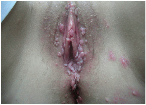
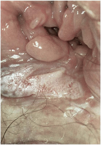
7

When clinical manifestations of vulvar HPV infection do occur, they often present as bumps or growths on the vulvovaginal or perianal epithelium. Commonly asymptomatic, genital warts are as likely to cause itching, burning, pain, or bleeding. On examination, warts can range in morphologic appearance from flat-topped papules to flesh-colored, dome-shaped papules, keratotic warts, or true condylomata acuminata (Fig. 14.2). External genital warts tend to occur on the moist surfaces of the vulva, introitus, and perianal area. Distinguishing warts from vulvar neoplasia based on appearance alone is not always possible. If the diagnosis is uncertain, or the patient is immunocompromised, biopsy should be undertaken.2

Women with genital warts should undergo routine cervical cancer screening. By the same token, the use of HPV testing, a change in the frequency of cervical cytologic screening or cervical colposcopy are not indicated in the presence of genital warts unless exophytic cervical warts or vulvar neoplasia is confirmed by biopsy; in which case, referral for colposcopic evaluation is indicated.2


FIGURE 14.2 Condyloma. (Multiple flat-topped papules and verrucous lesions.) (From C. K. Stockdale personal library.)

Spontaneous regression of genital warts occurs in up to 30% of affected patients. Despite this, many women opt for intervention to shorten the duration of lesions. It is important to note that regression does not necessarily lead to viral clearance because viral genomes can be detected in normal epithelium for months to years following clearing of visible disease. In immunocompetent women, however, the cell-mediated immunity that resulted in lesion regression most likely controls latent HPV infection. Disease recurrence is, therefore, less likely. Women at increased risk of developing HPV related manifestations thus include those receiving long-term corticosteroid therapy or chronic immunosuppressive treatment as well as immunosuppressed women with HIV infection. Antiviral chemotherapies are particularly critical for these populations if disease recurrences are to be minimized.8 However, most currently available treatment options are directed toward removal of visible disease rather than eradication of HPV infection.

Treatments endorsed by the Centers for Disease Control and Prevention include podofilox 0.5% solution or gel (patient-applied) as well as the provider-administered therapies: cryotherapy, podophyllin resin 10 to 25%, trichloroacetic or bichloroacetic acid 80 to 90%, surgical removal, and laser therapy.2 Such therapies have been the mainstay of treatment, along with imiquimod, a targeted antiviral therapy. Although most treatments are equivalent in terms of clearance rates, recurrence rates can be high, particularly for laser therapy where the recurrence rate exceeds 60%. However, imiquimod has been demonstrated to have significantly lower recurrence rates (9 to 19%).7

Intralesional interferon, a medication with antiproliferative, antiviral, and immunomodulatory properties, has, unlike imiquimod, demonstrated limited efficacy and is not recommended for first-line therapy in treating either warts or VIN.8 Its use, however, can be considered an alternative regimen for the treatment of external genital warts.2 The most recent FDA-approved patient-applied treatment for external condyloma is Veregen 15% topical ointment (Doak Dermatologics, Fairfield, NJ), a class of sinecatechins. This class of drugs is a mixture of catechins and other green tea components and has a response rate up to 55% complete clearance and 78% reduction of external anogenital warts with short-term follow-up.7

Because of decreased immune function, condyloma burden may exacerbate during pregnancy. Treatment is limited to expectant management or locally destructive options. Podofilox, podophyllin, and imiquimod are recognized teratogens and are therefore not recommended for use in pregnancy. Additionally, the risk of HPV appears inconsistent following vaginal delivery and does not justify routine cesarean birth in women with genital warts.2


Vulvar Intraepithelial Neoplasia

The most common symptom of VIN is pruritus. The incidence of VIN has increased over 400% since the 1970s especially among younger women.7 In 2004, the Vulvar Oncology Subcommittee of the International Society for the Study of Vulvar Diseases (ISSVD) developed the current classification system for VIN9 (Table 14.1). In this classification system, two distinct clinicopathologic subtypes of VIN exist: (a) VIN, usual type, which encompasses the former subcategories of VIN, warty type; VIN, basaloid type; and VIN, mixed (warty, basaloid) type; and (b) VIN, differentiated type, which encompasses the former category simplex type. Previously, VIN was categorized as VIN I, II, and III according to the degree of abnormality. In the new classification system, the term VIN is limited to histologically high-grade
squamous lesions (formerly termed VIN II and VIN III) for which treatment is indicated to prevent progression to cancer.


TABLE 14.1 Classification of Vulvar “Dystrophy” and Squamous Vulvar Intraepithelial Neoplasia


“Dystrophic” conditions


Lichen sclerosus


Squamous cell hyperplasia (hyperplastic “dystrophy”)


Mixed lichen sclerosus and squamous hyperplasia


Squamous vulvar intraepithelial neoplasia grade I (VIN I, low-grade VIN)


Squamous vulvar intraepithelial neoplasia using ISSVD 2004 modified terminology (includes only VIN II/III or high-grade VIN)


VIN, usual type


VIN, warty type


VIN, basaloid type


VIN, mixed (warty/basaloid) type


VIN, differentiated type (previously carcinoma in situ differentiated or simplex type)


VIN, vulvar intraepithelial neoplasia; ISSVD, International Society for the Study of Vulvar Disease.


Data from Sideri M, Jones RW, Wilkinson EJ, et al. Squamous vulvar intraepithelial neoplasia: 2004 modified terminology, ISSVD Vulvar Oncology Subcommittee. J Reprod Med. 2005;50(11):807-810; Stoler MH, Mills SE. The vulva and vagina. In: Mills SE, ed. Sternberg’s Diagnostic Surgical Pathology. 5th ed. Philadelphia: Lippincott Williams & Wilkins; 2010:2106.


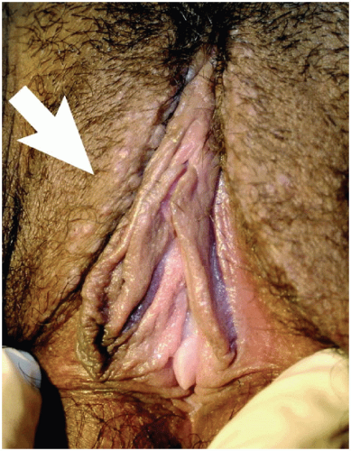
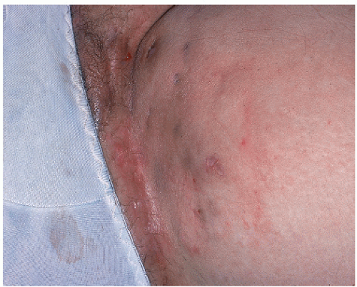
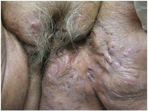
Usual type VIN, which includes warty-bowenoid or mixed type as well as pure warty and pure basaloid types, includes both VIN II and III, is HPV-related, commonly presents in women in their 30s and 40s, tends to be multifocal, and is strongly associated with cigarette smoking.10 Risk for usual type VIN is also increased in women who are immunosuppressed or immunodeficient.
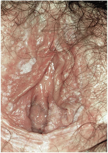
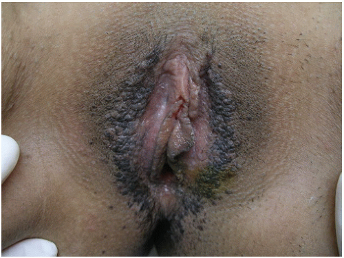
11 The interlabial grooves, posterior fourchette, and perineum are most frequently affected by multifocal lesions and more extensive disease is often confluent, involving the labia minora and majora as well as the perianal skin. Multifocal sites or confluence of usual type VIN is seen in up to 67% of women with usual VIN (Figs. 14.3, 14.4 through 14.5).

Usual type VIN is more common than differentiated VIN and accounts for at least 90% of cases of VIN. It is strongly HPV associated, with HPV-16 implicated in 70% of cases.12 Patients often have a history of condyloma, sexually transmitted infections, and HIV-positive status. Less than 10% of women with warty-bowenoid VIN develop invasive squamous cell carcinoma (SCC).
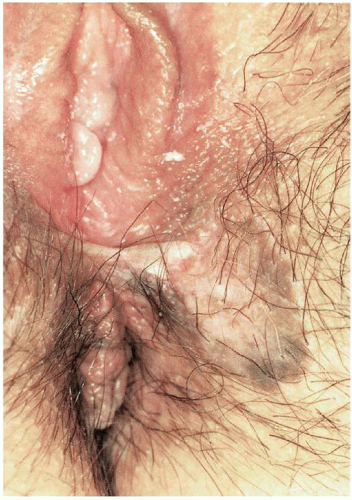
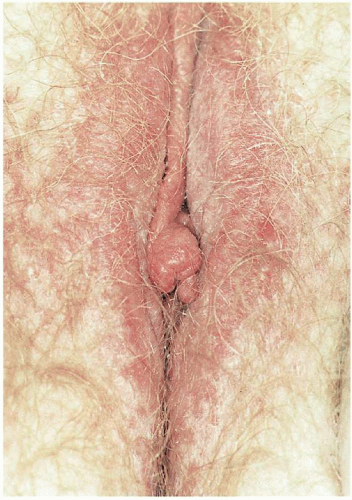
13 Younger women with multifocal, small, papular, pigmented lesions (a clinical entity referred to as bowenoid papulosis; Fig. 14.6) are the most likely to experience spontaneous resolution of the lesions, particularly if they are pregnant or postpartum at the time of diagnosis.14 In contrast, women older than 40 years of age and those who are immunosuppressed are at the greatest risk of progression.


FIGURE 14.3 Diffuse plaques of white epithelium in a 25-year-old woman with history of two prior laser treatments. Biopsies demonstrated VIN II (polymerase chain reaction was positive for HPV 16/18). (From Wilkinson EJ, Stone IK, eds. Atlas of Vulvar Disease. Philadelphia: Lippincott Williams & Wilkins; 2012:92.)


FIGURE 14.4 Hyperpigmented, localized lesion in an asymptomatic patient. Biopsy demonstrated VIN II/III, and the lesion was excised under general anesthesia and confirmation of diagnosis. Skin was undermined and closed with a permanent suture, which was removed 10 days later. (From Wilkinson EJ, Stone IK, eds. Atlas of Vulvar Disease. Philadelphia: Lippincott Williams & Wilkins; 2012:92.)

Differentiated VIN, which includes simplex VIN, is typically HPV negative, is seen most frequently in older women with other epithelial disorders such as lichen sclerosus or lichen simplex chronicus and is usually unifocal and unicentric. Although simplex VIN accounts for only 2 to 10% of biopsy specimens with VIN, it is more likely to progress to SCC than classic VIN. In one review looking at the natural history of differentiated VIN, 9% of untreated patients progressed to invasive vulvar carcinoma compared to 3.3% of treated patients.15,16 Differentiated VIN often occurs adjacent to invasive
SCC of the vulva and is thus regarded as the most common precursor to vulvar SCC. It may be often underdiagnosed.17 By definition, simplex VIN is a high-grade lesion and warrants further evaluation and treatment. In general, differentiated VIN lesions are smaller and less bulky than warty-baseloid VIN lesions, and often appear as gray-white lesions with roughened surfaces or as raised white plaques or nodules. It is often found during surveillance examinations for women with a history of lichen sclerosus or vulvar SCC. All suspicious lesions of the vulva should be biopsied.


FIGURE 14.5 Thickened white epithelium at introitus. Note prominent fissure. Excisional biopsy demonstrated VIN III. (From Wilkinson EJ, Stone IK, eds. Atlas of Vulvar Disease. Philadelphia: Lippincott Williams & Wilkins; 2012:93.)


FIGURE 14.6 Vulvar intraepithelial neoplasia III. (Warty-bowenoid VIN; diffuse brown hyperpigmented raised papules.) (From C. K. Stockdale personal library.)

VIN is highly variable in its gross appearance; because the lesions may be subclinical, they may only be apparent with colposcopy. Because the prevalence of multifocal lesions is high, comprehensive colposcopic examination of the entire lower genital tract and perianal area is indicated in any woman with VIN. The most common findings noted on the vulva during examination with the colposcope are areas of acetowhitening that may or may not have areas of punctation. When using colposcopy to evaluate for VIN, it is important to remember that the vulvar area must be soaked with 2 to 5% acetic acid for about 5 minutes. Acetowhite changes do not determine the diagnosis; rather, they are nonspecific and only serve to indicate where biopsies should be taken. Raised acetowhite areas, particularly in conjunction with small papillations or satellite lesions, suggest HPV changes. Lesions that are hyperpigmented, fixed, indurated, or ulcerated, as well as condylomas that do not respond to therapy warrant biopsy evaluation. Vulvar punch biopsies are usually done with the Keys dermatologic biopsy instrument because it allows for full-thickness evaluation of the vulva skin as well as accurate orientation of the specimen. Multiple biopsies may be required when there are multifocal lesions. Bleeding from the biopsy site is usually controlled with ferric subsulfate (Monsel solution) or silver nitrate sticks; rarely are sutures necessary. If a punch biopsy does not yield a satisfactory specimen or if the lesions are quite indurated or have extensive ulceration, an excisional biopsy may be needed. If melanoma is suspected, excisional biopsy is indicated. Any lesion on the vulva that is not known to be benign warrants a biopsy, and if there are multiple areas of abnormalities, multiple biopsies are indicated.

The goals of treatment for VIN are to prevent the development of invasive vulvar cancer and alleviate symptoms, without marked changes in the normal anatomy and function of the vulva. Treatment is individualized, with consideration of the biopsy results, location and extent of the disease, and the patient’s symptoms. Observation suffices for VIN I. For patients with VIN II or VIN III, treatment usually is recommended, although observation is initially reasonable for women who have recently completed a course of corticosteroids, who are temporarily immunocompromised, or who are pregnant.


Management options currently include wide local excision, skinning vulvectomy, laser ablation, and topical treatment. Wide local excision or laser therapy for patients with no evidence of gross or microscopic invasion remains the primary treatment used for VIN. The surgical approaches for treatment appear to be similarly effective.16 Treatment should go to a depth of 1 to 2 mm in non-hair-bearing areas and may need to extend to 3 mm in hair-bearing areas of the vulva in order to encompass any disease around the hair follicle. Disease-free margins of at least 1 cm should be excised around the lesion.

Topical agents that enhance or induce strong cell-mediated immune responses likely hold the greatest promise not only for control of HPV-related disease but also for reduction of future recurrences. Prior to the use of topical treatments, a careful colposcopic examination with biopsies must be done to rule out invasive disease. Topical 5% imiquimod acts by activating macrophages and dendritic cells to release interferon and other proinflammatory cytokines. These cytokines in turn lead to activation of the appropriate antigen-specific immune response. Imiquimod efficacy in the treatment of genital warts is well documented, and in a number of recent trials, its use has been associated with encouraging results in the treatment of VIN II and VIN III with a response rate approaching 90%.15,18,19 Furthermore, van Seters found regression of VIN was associated with clearance of HPV.19 Imiquimod has been associated with relief of pruritus and pain both immediately and for up to 12 months after use.19 Side effects of imiquimod are usually local and may include mild to moderate erythema, erosion, burning, and ulceration.

Current treatments for VIN, although numerous, have largely resulted in suboptimal outcomes. Response rates have been poor and relapse rates high. Additionally, the resultant physical and psychological morbidity associated with treatment can be profound.

At least one-third of patients develop recurrent VIN regardless of the prior treatment modality. Risk factors for recurrence include immunosuppression, multifocal or multicentric disease, and positive margins on the biopsy specimen. For this reason, patients treated for VIN should be followed closely every 3 to 4 months until they are disease free for at least 2 years.


Vaginal Intraepithelial Neoplasm

Although the true incidence of vaginal intraepithelial neoplasm (VAIN) is not known, the diagnosis has been increasing over the last several years due to increased awareness, expanded cytologic screening, and more liberal use of colposcopy. The average patient is between 43 and 60 years of age. Infection with HPV is a common association.

VAIN is the terminology used to describe vaginal dysplasia. This spans the spectrum from VAIN I and II (only the lower one-third and two-thirds of the epithelium, respectively, is involved) to VAIN III (more than two-thirds of the epithelium is involved). Full-thickness involvement of the epithelium, or carcinoma in situ, is included under VAIN III. VAIN is consistently associated with prior or concurrent neoplasia elsewhere in the lower genital tract.

Premalignant neoplasms are almost always asymptomatic and are often detected by an abnormal Pap smear, although some patients may present with postcoital spotting or vaginal discharge. A tissue diagnosis must be established when an abnormal cytologic smear of the vaginal cuff is obtained. Colposcopy is performed with biopsy of the most abnormal area. If there is any question of invasion, excision under anesthesia must be undertaken. In postmenopausal patients, a few weeks of topical estrogen may help visualization and improve detection of VAIN.

After application of acetic acid, lesions often appear as raised or flat white, granular epithelium with well-defined borders; punctuation may or may not be present. Biopsies should be taken of any abnormal appearing areas. It may be helpful to slightly close the speculum prior to biopsy so the vaginal wall is not taut with tension, which makes performing a biopsy difficult. Local anesthetic (without epinephrine) should be used prior to biopsy. The choice of biopsy instrument depends on the type and location of the lesion. Sessile lesions may usually be biopsied with cervical biopsy forceps, whereas some raised lesions may be grasped with a forceps and affine scissors maybe sued for biopsy. The majority of lesions are in the upper one-third of the vagina, so in patients posthysterectomy, they may be difficult to see and skin hooks may be needed to evert areas of vaginal wall recess. If the surface is irregular or there are severe vascular abnormalities, an excisional biopsy should be taken to exclude an invasive process. Hemostasis can usually be obtained with ferric subsulfate (Monsel solution) or silver nitrate; in some cases, a suture may be necessary.

A variety of treatment options are available for VAIN: excision, ablation, topical chemotherapy, and radiation. Management should be individualized with consideration given to the presence of multifocal disease, any history of previous treatment failures, the woman’s general health and risks from surgery, her desire to preserve her sexual function, and the degree to which the provider is sure that invasive disease has been ruled out. Some manage VAIN I with close surveillance. In general, surgical excision is the most common form of VAIN treatment. This may require local excision, partial vaginectomy, and rarely, total vaginectomy. Ablation is most commonly done with the CO2 laser, and up to one-third of patients may need more than one treatment. There are no guidelines clearly defining the ideal topical treatment for multifocal high-grade VAIN, although it appears to
be appropriate first-line therapy for women with early lesions and multifocal disease or those who are poor surgical candidates. Imiquimod and 5-fluorouracil are the topical agents most commonly used. Radiation therapy is reserved for patients who have failed previous treatments for VAIN; are poor surgical candidates; or have extensive, multifocal disease. Because VAIN is usually multifocal, follow-up every 6 months for up to 2 years and then annually thereafter is warranted.


Extramammary Paget Disease

Extramammary Paget disease is an uncommon lesion that arises in skin rich in apocrine glands, with limited information based on case reports and case series owning to its rarity. Vulvar Paget disease is most often an apocrine carcinoma in situ. However, 4% of women will have a contiguous adenocarcinoma and up to 26% will have noncontiguous underlying adenocarcinoma.20,21 Patients characteristically present with symptoms including burning or itching, with a reported age range of 47 to 93 years in one case series.
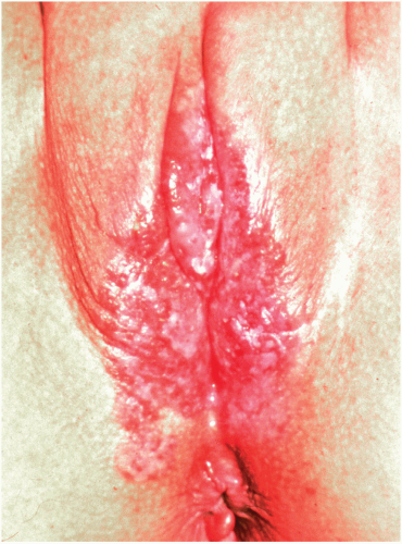
22 The classic appearance of vulvar Paget disease is a red velvety plaque, which may be mistaken for vulvar eczema (Fig. 14.7). It is usually multifocal and may occur anywhere on the vulva, mons, perineum/perianal area, or inner thigh. Vulvar biopsy should be performed in patients with suspicious lesions, including those with persistent pruritic eczematous lesions that fail to resolve within 6 weeks of appropriate antieczema therapy.


FIGURE 14.7 Paget disease of the vulva. The lesion is red, moist, and sharply demarcated. (From Rubin E, Farber JL. Pathology. 3rd ed. Philadelphia: Lippincott Williams & Wilkins; 1999.)

Paget disease is diagnosed by histologic appearance including the presence of signet-ring or Paget cells. Following biopsy confirmation of vulvar Paget disease, patients should be screened for possible associated or synchronous cancers with pelvic exam, cervical cytology, endometrial biopsy, and mammography, as well as targeted evaluation for anal (occult blood testing, endoscopy) or urethral (urine cytology, cystoscopy) lesion involvement given the risk for underlying adenocarcinoma.22

Treatment of noninvasive Paget disease is classically by surgical excision with limited evidence for nonexcisional treatment modalities such as laser ablation, radiotherapy, photodynamic therapy, and imiquimod. The risk of recurrence following excision is reported to occur in up to 60% of women regardless of margin status or extent of surgery and is attributed to the presence of “skip lesions.”22 The use of frozen section to ensure complete excision (negative margin status) has been employed to reduce the high rate of recurrence without clear benefit. Given the risk of recurrence and progression to invasive disease, long-term monitoring with biopsy of any abnormal vulvar lesion is paramount in patients with a history of Paget disease.


VULVAR DERMATITIS

The term dermatitis (or eczema) describes a poorly demarcated, erythematous, and usually itchy rash. Burning can sometimes occur in cases of mucosal involvement. Subtypes are numerous and can be classified as either exogenous (irritant or allergic contact dermatitis) or endogenous (atopic dermatitis). Endogenous or atopic dermatitis has a familial predisposition and may begin in childhood.

Dermatitis is common and has been reported to occur in 20 to over 60% of patients seen with chronic vulvar symptoms, with atopic dermatitis being by far the most frequently encountered.23 Unfortunately, the clinical appearance of the vulva often does not help secure the diagnosis, and in many cases, more than one process has led to the symptoms the patient reports. It is not uncommon to see a mixed picture where endogenous dermatitis or another epithelial disorder has been worsened by use of ointments or creams to which the patient has adversely reacted.23,24 Vulvar dermatitis often presents as chronic irritation and/or pruritus (that is often nocturnal), and patients report persistent rubbing and itching to alleviate the symptoms. The symptoms may be worsened by stress, menstruation, heat, or sweat. Chronic scratching or rubbing by the patient may lead
to histologic changes in the dermis referred to as squamous hyperplasia or lichen simplex chronicus.


Allergens, Irritants, and Contact Dermatitis

Contact dermatitis is one of the most frequently encountered and often simultaneously avoidable problems seen in vulvar specialty clinics with a reported prevalence of up to 54% in vulvar clinic patients.25 The susceptibility of the vulvar skin to inflammation is widely underestimated by patients as well as providers. Compared to other areas of exposed skin, the stratum corneum of the vulva functions less efficiently as a barrier, which increases its susceptibility to irritants. Many seemingly innocuous behaviors, such as bathing, the use of sanitary or incontinence pads, or exposure to known irritants or allergens in numerous topical medications, have the potential to initiate this eczematous process.24 Active and inert components of topical medications are a very common cause of allergic vulvar dermatitis. Latex sensitivity or reactions to proteins in seminal plasma are infrequent causes.26 It is important to remember that a patient may have used an agent for months or years without any problem and then have an “allergy” develop. Additionally, self-treatment (using over-the-counter agents or alternative therapies) often exacerbate the process prior to medical evaluation.

Irritant contact dermatitis has been identified in up to 26% of women diagnosed with vulvar dermatitis, often as a result of exposure to such irritants as detergents; soaps; perfumes; semen; and propylene glycol, an additive found in many topical medications. Allergic contact dermatitis, in contrast, is an immunologically mediated inflammatory reaction (type IV delayed hypersensitivity) to an allergen in a previously sensitized woman. Although distinguishing allergic from irritant contact dermatitis can be difficult, the intermittent nature of the symptoms and the timing of symptom onset (10 to 14 days following exposure but may be less than 24 hours if already sensitized) serve as important keys to making the diagnosis of the former. The list of commonly encountered allergens, although similar to that of common irritants, also includes a variety of topical medications such as antibiotics (e.g., neomycin, bacitracin), antifungals (clotrimazole, miconazole), all corticosteroids, and the greatest offenders, the topical anesthetics.24 Additionally, close questioning of the patient’s personal practices is an important way to detect potential irritants and/or allergens as well as unhealthy vulvar hygiene habits. In one study, over 60% of women attending a specialty clinic for vulvar disease described personal hygiene and/or self-treatment practices that may contribute to vulvar dermatitis.26

The presence of contact hypersensitivity in an underlying vulvar dermatitis should be suspected in cases where a patient does not respond following therapy or experiences a recurrence of symptoms after initial remission.25 In this case, patch testing may be beneficial. Not only will patch testing help to distinguish between atopic and contact dermatitis, it may help determine the cause of the dermatitis. There is a standard patch test series in the United States that tests for the most common and important allergens in North America. Positive patch tests for relevant substances occur in 25 to 60% of women with vulvar pruritus.27

On examination, clinical signs can range from mild erythema, swelling, and scaling to marked erythema, fissures, edema, and vesicles or bullae. Chronic eczema is often erythematous and scaly (Fig. 14.8). Secondary infection may be present as well. Confirmation of the diagnosis should include assessment to rule out candidiasis. Biopsy, which will show nonspecific changes such as spongiosis, parakeratosis, and some dermal inflammatory infiltrate, is not helpful.

Treatment consists of removal of the offending agent or practice, correction of barrier function, elimination of scratching, and reduction of inflammation. Barrier function should be restored through the use of Sitz baths, estrogen therapy if indicated, treatment of concomitant
infection if present, and application of a thin layer of plain petrolatum. Exposing the vulva to warm water, without any additives, for at least 5 minutes in the morning and again at night, helps to rehydrate the skin and soothe the sensory nerve endings of the vulva. The moisture may be sealed in with a topical corticosteroid ointment. Medications with antihistamine and sedative properties, such as doxepin or hydroxyzine (10 to 50 mg in the evening), can be added to control nocturnal itching, whereas a selective serotonin reuptake inhibitor (SSRI) can be used during the day. Nonsedating antihistamines do not help with vulvar pruritus. Anti-inflammatory therapy should also begin with a mid- to high-potency corticosteroid for 2 to 3 weeks (Table 14.2). A weaker corticosteroid, such as 1% hydrocortisone, can then be continued as needed. There is no evidence that application of topical corticosteroids more than once daily is beneficial.28


FIGURE 14.8 Moderate erythema in patient who complains of pruritus for 3 years. Clinical impression was eczema, and patient was advised to use hypoallergenic soap. Condition resolved. (From Wilkinson EJ, Stone IK, eds. Atlas of Vulvar Disease. Baltimore: Lippincott Williams & Wilkins; 1995:81, with permission.)

In recalcitrant cases, oral or intramuscular (IM) corticosteroids may be necessary.24 IM triamcinolone 60 mg may be given IM every 6 weeks for up to three doses. After the first injection, petrolatum jelly may be used twice a day after sitz baths for 2 weeks followed by use of a superpotent topical corticosteroid. Use of IM triamcinolone may cause hyperglycemia, changes in menstruation, and adrenal suppression. It should not be used in women with a history of either severe depression or bipolar disease.

An intralesional injection of triamcinolone acetonide 3.3 to 10 mg/mL may also be tried in resistant cases. A total of 1 to 2 mL is given by injecting small amounts throughout the entire lesion or plaque; this may be repeated monthly up to three times. It may be helpful to apply a topical anesthetic such as EMLA prior to injection.


Lichen Simplex Chronicus

Vulvar lichen simplex chronicus, also known as squamous hyperplasia, is a common, chronic eczematous disease characterized by intense and unrelenting itching and scratching. In vulvar specialty clinics, lichen simplex chronicus accounts for 10 to 35% of patients evaluated. Although lichen simplex chronicus occurs primarily in middle to late adult life, it can occur in children. Up to 65 to 75% of patients will report a history of atopic disease (hay fever, asthma, childhood eczema), and as such, lichen simplex chronicus can be seen as a localized variant of atopic dermatitis. Patients often present with intractable itching and pruritus, and most will admit to vigorous scratching or rubbing. The pruritus is often worse at night, and chronic scratching may lead to lichenification, fissures, and postinflammatory pigment changes. Thus, lichen simplex chronicus represents an end-stage response to a wide variety of possible initiating processes, including environmental factors (e.g., heat, excessive sweating, and irritation from clothing or topically applied products) and dermatologic disease (e.g., candidiasis, lichen sclerosus).29,30


TABLE 14.2 Common Topical Steroids


Topical Steroid Class


Name (Trade)


Name (Generic)


Common Uses


Class I Super-potent


Temovate (0.05% cream, ointment)a Ultravate (0.05% cream, ointment)


Clobetasol propionate Halobetasol propionate


Lichen sclerosus Lichen planus


Class II High potency


Lidex (0.05%)


Fluocinonide


Psoriasis Lichen simplex chronicus


Class III


Valisone (0.1% ointment)


Betamethasone valerate


Class IV Midpotency


Kenalog (0.1% cream) Westcort (0.2% ointment)


Triamcinolone Hydrocortisone valerate


Eczema Mild irritation


Class V


Locoid (0.1% cream) Valisone (0.1% cream) Westcort (0.2% cream)


Hydrocortisone butyrate Betamethasone valerate Hydrocortisone valerate


Class VII Mild


Hydrocortisone 1.0%, 2.5%


Hydrocortisone


Mild eczema Maintenance


a Creams tend to work better in the vagina due to better absorption. Ointments work well on the vulva because they are less irritating than creams, solutions, jelly, or lotions and provide a soothing barrier.


Data from Ference JD, Last AR. Choosing topical corticosteroids. Am Fam Physician. 2009;79(2):135-140.


Clinically, lichen simplex chronicus appears as one or more erythematous, scaling, lichenified plaques (Fig. 14.9). Various degrees of excoriation are often visible. In long-standing disease, the skin appears thickened and leathery, and areas of hyperpigmentation and hypopigmentation may be present. Erosions and ulcers can also develop, most commonly from chronic scratching. Vaginal fungal cultures are helpful in determining the presence of an underlying condition on which lichen simplex chronicus is superimposed. Biopsy (which would demonstrate marked hyperkeratosis with widening and deepening of the rete ridges) is rarely necessary unless an underlying disease (such as lichen sclerosus or psoriasis) is suspected or the patient fails to respond to treatment.

Treatment consists of identification and removal of the initiating factor, repair of the skin’s barrier layer function, reduction of inflammation, and disruption of
the itch-scratch cycle. Mid- to high-potency topical corticosteroids (depending on the presence of underlying disease) should be applied nightly until symptoms begin to abate; less frequent use (e.g., alternate nights, then twice weekly) should continue until the condition resolves. As noted previously, medications with antihistamine and sedative properties can be added to control nocturnal itching; an SSRI for daytime use and oral or intralesional steroids can be used for refractory cases. Given the side effects of local steroid therapy, calcineurin inhibitors tacrolimus (0.03% ointment) and pimecrolimus (1% cream) may be considered in refractory cases and have been approved as second-line anti-inflammatory agents for the treatment of dermatitis.31 These are used sparingly twice a day for 14 to 30 days followed by maintenance therapy twice a week. Upon discontinuation, the dermatitis recurs in 35 to 54% of patients. The use of tacrolimus may cause burning or stinging and these side effects may be minimized through the application of a film of petrolatum jelly prior to use of the ointment.


FIGURE 14.9 Mild lichen simplex chronicus. Note the subtle lichenification (thickening) on the labia majora bilaterally (arrow). (From Boardman LA, Botte J, Kennedy CM. Recurrent vulvar itching. Obstet Gynecol. 2005;105[6]: 1451-1455.)


Seborrheic Dermatitis

Seborrheic dermatitis is very rare on the vulva, and its etiology is not known. It usually affects the scalp, face, presternal, and intrascapular areas (areas rich in sebaceous glands). Lesions are pale to yellowish red, with greasy, nonadherent scales and are not clearly defined. Secondary infection may occur. Vulvar lesions are usually distributed symmetrically on the mons pubis and genitocrural folds where sebaceous glands are concentrated; involvement of labia minora is rare.

Clinically, seborrhea may be confused with psoriasis, candidiasis, tinea cruris, or lichen simplex chronicus. The diagnosis is one of exclusion. When present in the genital region, there will often be seborrheic dermatitis involving other areas of the body.

Initial treatment consists of low- to high-potency corticosteroid ointment as a burst and taper with or without a topical antifungal such as ketoconazole.29 If topical steroids are not adequate in controlling symptoms, further treatment considerations may include selenium sulfide, salicylic acid, and tar shampoo. However, the use of these agents may be limited by contact irritation to sensitive vulvar skin.


Hidradenitis Suppurativa

Hidradenitis suppurativa, also termed acne inversa and Verneuil disease, is a common chronic, suppurative, cutaneous process that results from occlusion of follicles. It commonly involves the intertriginous skin of the axillary, groin, perineal, perianal, and inframammary regions (Figs. 14.10 and 14.11). Scarring of the affected sites results from chronic infection and draining abscesses.

The prevalence of hidradenitis suppurativa is estimated to be as high as 4%, although hidradenitis suppurativa is often underrecognized and underdiagnosed.32,33 Women are more likely to be affected than men by a ratio of about 3:1. Smoking seems to be a major triggering factor. Other causal factors may include both acquired and genetic characteristics including onset after puberty and prior to age 40 years, obesity (with hyperandrogenism),
association with other endocrine disorders (e.g., diabetes), and an apparent familial predisposition in some patients. Axillary and inguinal involvement is more common in females, whereas perianal and buttocks involvement is more common in males.33


FIGURE 14.10 Hidradenitis suppurativa. This patient has involvement of the inguinal areas, labia majora, and mesial thighs. Her undergarment was stained by the oozing lesions. (From Goodheart HP, MD. Goodheart’s Photoguide of Common Skin Disorders. 2nd ed. Philadelphia: Lippincott Williams & Wilkins; 2003.)


FIGURE 14.11 Hidradenitis suppurativa. (Note scarring, pitting, and multiple fistulous tracts.) (From C. K. Stockdale personal library.)

The onset of hidradenitis suppurativa is insidious. Early diagnosis of hidradenitis suppurativa is easily overlooked and often attributed to simple comedones of acne. However, unlike acne, significant sebaceous gland involvement is not a component of hidradenitis suppurativa. Early symptoms consist of pruritus, erythema, burning, and local hyperhidrosis.34 Occlusion of a hair follicle results in a cyst, similar to the comedones of acne. The follicular duct expands and eventually ruptures, which release a number of antigens and other inflammatory stimuli that cause the development of an acute inflammatory response in the surrounding tissue. Epithelial cells from the follicular wall, as well as stem cells, are released during the rupture as well and may promote cellular proliferation. The combination of acute inflammation and cellular proliferation is thought to cause the formation and spread of dermal and subcutaneous abscesses and sinus tracts that are seen with hidradenitis suppurativa. Lesions are extremely painful, and the average duration of a lesion is 7 days. Most patients have at least one active lesion at any given time. As the area heals, it becomes scarred and fibrotic. Chronic lymphedema may develop as a result of lymphatic destruction. Ultimately, hyperpigmentation, scarring, pitting, and multiple fistulous sites are noted.

Although specific diagnostic criteria do not exist, the diagnosis becomes apparent with disease progression; biopsy is neither required nor diagnostic. Clinical criteria suggesting hidradenitis suppurativa include recurrent deep boils for more than 6 months in flexural apocrine gland skin sites (typically involving the axilla, inguinal, and anogenital areas), onset after puberty, poor response to conventional antibiotics, and tendency toward relapse and recurrence.35 Late disease is characterized by chronic infection, draining abscesses, nodules, sinus tracts, scarring, and hyperpigmentation. Hidradenitis is graded according to Hurley’s clinical staging system or the more recently developed modified Hidradenitis Suppurativa Score (Tables 14.3 and 14.4). The severity of hidradenitis suppurativa can be classified according to the Hurley’s stage, which correlates with the extent of
treatment that is likely to be needed. The modified score includes the anatomic region affected by the disease, the number and characteristics of lesions, the longest distance between two relevant lesions, and the presence of normal skin bridges between lesions.


TABLE 14.3 Hurley’s Clinical Staging


The severity of HS can be classified according to the Hurley’s stage, which correlates with the extent of treatment that is likely to be needed.


  • Stage I—Abscess formation, single or multiple, without sinus tracts and cicatrization (scarring). Although classically managed with drug therapy, hormonal and metabolic controls are now part of the treatment.


  • Stage II—Recurrent abscesses with tract formation and cicatrization. Single or multiple widely separated lesions. Managed with drug therapy and limited excision of recalcitrant lesions. Unroofing is emerging as the preferred technique.


  • Stage III—Diffuse or near diffuse involvement, or multiple interconnected tracts and abscesses across the entire area. Unlikely to benefit from medical therapy alone; wide surgical excision (either unroofing or en bloc excision) is recommended either as adjunctive or curative therapy.


HS, hidradenitis suppurativa.


From Hurley, HJ. Axillary hyperhidrosis, apocrine bromhidrosis, hidradenitis suppurativa, and familial benign pemphigus: surgical approach. In: Roenigk RK, Roenigk HH, eds. Dermatologic Surgery. New York: Marcel Dekker; 1989:729.


TABLE 14.4 Modified Hidradenitis Suppurativa Score


Characteristics of Hidradenitis Suppurativa Lesions


Points per Lesion


Anatomic region


Any


3


None


0


Types of lesions


Fistula


4


Nodule


2


Abscess


1


Scar


1


Other


1


Total area involveda


<5 cm


2


5-10 cm


4


>10 cm


8


Are all lesions clearly separated by normal skin?


Yes


0


No


6


Total score:


The Hidradenitis Suppurative Score (HSS) as modified by Sartorius was proposed to standardize comparative analyses of treatment for HS.


a Record the longest distance between two relevant lesions (e.g., nodule and fistula) in each affected region, or record the size of a single lesion in each affected region.


Data from Sartorius K, Lapins J, Emtestam L, et al. Suggestions for uniform outcome variables when reporting treatment effects in hidradenitis suppurativa. Br J Dermatol. 2003;149:211; Mahmoud BH, Tierney E, Hexsel CL, et al. Prospective controlled clinical and histopathologic study of hidradenitis suppurativa treated with the long-pulsed neodymium:yttrium-aluminium-garnet laser. J Am Acad Dermatol. 2010;62:637-645.


The differential diagnosis of hidradenitis suppurativa includes Crohn disease, follicular pyoderma, acne vulgaris, and Fox-Fordyce disease. Finally, SCC arising in hidradenitis suppurativa is rare but has been reported. Thus, although biopsy is not indicated for the diagnosis of hidradenitis suppurativa, it is indicated when there is concern for malignancy.

Treatment of hidradenitis suppurativa is challenging and based primarily on anecdotal evidence (Table 14.5). Patients need to be assured that the disease is not contagious nor is it due to poor hygiene. Current treatment is directed at prevention of new lesions through medical management and elimination of existing lesions surgically when appropriate. Smoking cessation is imperative. In order to improve the local environment, avoidance of irritants, heat, sweating, and reducing friction (e.g., avoidance of tight clothing, limit pad use) is encouraged. Additionally, weight reduction is often helpful. The use of washcloths should be discouraged as should high-pH soaps, whereas the use of clean bare hands for cleansing affected areas and neutral-pH detergents may be encouraged. Overall, treatments including oral antibiotics, topical clindamycin, isotretinoin, systemic and intralesional corticosteroids, and surgery have been inconsistently effective. There is no data to support the use of one medical therapeutic approach over another.

Only gold members can continue reading. Log In or Register to continue

Stay updated, free articles. Join our Telegram channel

Jun 25, 2016 | Posted by in GYNECOLOGY | Comments Off on Benign Disorders of the Vulva and Vagina

Full access? Get Clinical Tree

Get Clinical Tree app for offline access