Chapter 201 Bartonella
The spectrum of disease resulting from human infection with Bartonella species includes the association of bacillary angiomatosis and cat-scratch disease (CSD) with Bartonella henselae. Six major Bartonella species are pathogenic for humans: B. henselae, Bartonella quintana, Bartonella bacilliformis, Bartonella elizabethae, Bartonella vinsonii, and Bartonella clarridgeiae (Table 201-1). Several other Bartonella species have been found in animals, particularly rodents and moles.
Members of the genus Bartonella are gram-negative, oxidase-negative, fastidious aerobic rods that ferment no carbohydrates. B. bacilliformis is the only species that is motile, achieving motility by means of polar flagella. Optimal growth is obtained on fresh media containing 5% or more sheep or horse blood in the presence of 5% carbon dioxide. The use of lysis centrifugation for specimens from blood on chocolate agar for extended periods (2-6 wk) enhances recovery.
201.1 Bartonellosis (Bartonella bacilliformis)
The 1st human Bartonella infection described was bartonellosis, a geographically distinct disease caused by B. bacilliformis. There are 2 predominant forms of illness due to B. bacilliformis: Oroya fever, a severe, febrile hemolytic anemia, and verruca peruana (verruga peruana), an eruption of hemangioma-like lesions. B. bacilliformis also causes asymptomatic infection. Bartonellosis is also called Carrión disease in honor of the Peruvian medical student who inoculated himself with blood from a verruca and 21 days later had Oroya fever. He died 39 days after the inoculation, thus proving the unitary etiology of the 2 clinical illnesses.
Etiology
B. bacilliformis is a small, motile, gram-negative organism with a brush of 10 or more unipolar flagella, which appear to be important components for invasiveness. An obligate aerobe, it grows best at 28°C in semisolid nutrient agar containing rabbit serum and hemoglobin.
Epidemiology
Bartonellosis is a zoonosis found only in mountain valleys of the Andes Mountains in Peru, Ecuador, Colombia, Chile, and Bolivia at altitudes and environmental conditions favorable for the vector, which is the sandfly, Lutzomyia verrucarum.
Pathogenesis
After the sandfly bite, Bartonella organisms enter the endothelial cells of blood vessels, where they proliferate. Found throughout the reticuloendothelial system, they then re-enter the bloodstream and parasitize erythrocytes. They bind on the cells, deform the membranes, and then enter intracellular vacuoles. The resultant hemolytic anemia may involve as many as 90% of circulating erythrocytes. Patients who survive this acute phase may or may not experience the cutaneous manifestations, which are nodular hemangiomatous lesions or verrucae ranging in size from a few millimeters to several centimeters.
Clinical Manifestations
The incubation period is 2-14 wk. Patients may be totally asymptomatic or may have nonspecific symptoms such as headache and malaise without anemia.
Oroya fever is characterized by fever with rapid development of anemia. Clouding of the sensorium and delirium are common symptoms and may progress to overt psychosis. Physical examination demonstrates signs of severe hemolytic anemia, including icterus and pallor, sometimes in association with generalized lymphadenopathy.
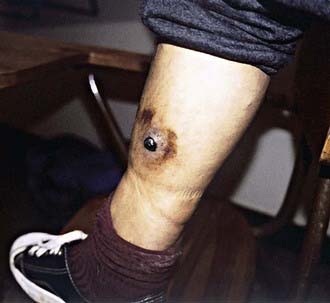
In the pre-eruptive stage of verruca peruana (Fig. 201-1), patients may complain of arthralgias, myalgias, and paresthesias. Inflammatory reactions such as phlebitis, pleuritis, erythema nodosum, and encephalitis may develop. The appearance of verrucae is pathognomonic of the eruptive phase. Lesions vary greatly in size and number.

Figure 201-1 A single large lesion of verruca peruana on the leg of an inhabitant of the Peruvian Andes. Such lesions are prone to superficial ulceration, and their vascular nature may lead to copious bleeding. Ecchymosis of the skin surrounding the lesion is also evident.
(Courtesy of Dr. J.M. Crutcher, Oklahoma State Department of Health, Oklahoma City.)
Diagnosis
The diagnosis is established on clinical grounds in conjunction with a blood smear demonstrating organisms or with blood culture. The anemia is macrocytic and hypochromic, with reticulocyte counts as high as 50%. B. bacilliformis may be seen on Giemsa stain preparation as red-violet rods in the erythrocytes. In the recovery phase, organisms change to a more coccoid form and disappear from the blood. In the absence of anemia, the diagnosis depends on blood cultures. In the eruptive phase, the typical verruca confirms the diagnosis. Antibody testing has been used to document infection.
Treatment
B. bacilliformis is sensitive to many antibiotics, including rifampin, tetracycline, and chloramphenicol. Treatment is very effective in rapidly diminishing fever and eradicating the organism from the blood. Chloramphenicol (50-75 mg/kg/day) has been considered the drug of choice, because it is also useful in the treatment of concomitant infections such as Salmonella. Fluoroquinolones have been used successfully as well. Blood transfusions and supportive care are critical in patients with severe anemia. Antimicrobial treatment for verruca peruana is considered when there are >10 cutaneous lesions, if the lesions are erythematous or violaceous, or if the onset of the lesions was <1 mo before presentation. Oral rifampin is effective in the healing of lesions. Surgical excision may be needed for lesions that are large and disfiguring or that interfere with function.
201.2 Cat-Scratch Disease (Bartonella henselae)
The most common presentation of Bartonella infection is CSD, which is a subacute, regional lymphadenitis caused by B. henselae. It is the most common cause of chronic lymphadenitis that persists for >3 wk.
Etiology
B. henselae can be cultured from the blood of healthy cats. B. henselae organisms are the small pleomorphic gram-negative bacilli visualized with Warthin-Starry stain in affected lymph nodes from patients with CSD. Development of serologic tests that showed prevalence of antibodies in 84-100% of cases of CSD, culturing of B. henselae from CSD nodes, and detection of B. henselae by polymerase chain reaction (PCR) in the majority of lymph node samples and pus from patients with CSD confirmed the organism as the cause of CSD. Occasional cases of CSD may be caused by other organisms; 1 report described a veterinarian with CSD caused by B. clarridgeiae.
Epidemiology
CSD is common, with more than 24,000 estimated cases per year in the USA. It is transmitted by cutaneous inoculation. Most (87-99%) patients have had contact with cats, many of which are kittens <6 mo of age, and >50% have a definite history of a cat scratch or bite. Cats have high-level Bartonella bacteremia for months without any clinical symptoms; kittens are more frequently bacteremic than adult cats. Transmission between cats occurs via the cat flea, Ctenocephalides felis. In temperate zones, the majority of cases occur between September and March, perhaps in relation to the seasonal breeding of domestic cats or to the close proximity of family pets in the fall and winter. In tropical zones, there is no seasonal prevalence. Distribution is worldwide, and infection occurs in all races.
Cat scratches appear to be more common among children, and boys are affected more often than girls. CSD is a sporadic illness; usually only 1 family member is affected, even though many siblings play with the same kitten. However, clusters do occur, with family cases within weeks of one another. Anecdotal reports have implicated other sources, such as dog scratches, wood splinters, fishhooks, cactus spines, and porcupine quills.
Pathogenesis
The pathologic findings in the primary inoculation papule and affected lymph nodes are similar. Both show a central avascular necrotic area with surrounding lymphocytes, giant cells, and histiocytes. Three stages of involvement occur in affected nodes, sometimes simultaneously in the same node. The first stage consists of generalized enlargement with thickening of the cortex and hypertrophy of the germinal center and with a predominance of lymphocytes. Epithelioid granulomas with Langhans giant cells are scattered throughout the node. The middle stage is characterized by granulomas that increase in density, fuse, and become infiltrated with polymorphonuclear leukocytes, with beginning central necrosis. In the final stage, necrosis progresses with formation of large pus-filled sinuses. This purulent material may rupture into surrounding tissue. Similar granulomas have been found in the liver, spleen, and osteolytic lesions of bone when those organs are involved.
Clinical Manifestations
After an incubation period of 7-12 days (range 3-30 days), 1 or more 3- to 5-mm red papules develop at the site of cutaneous inoculation, often reflecting a linear cat scratch. These lesions are often overlooked because of their small size but are found in at least 65% of patients when careful examination is performed (Fig. 201-2

Stay updated, free articles. Join our Telegram channel

Full access? Get Clinical Tree