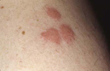
The type of reaction that occurs after an arthropod bite depends on the species of insect and the age group and reactivity of the human host. Arthropods may cause injury to a host by various mechanisms, including mechanical trauma, such as the lacerating bite of a tsetse fly; invasion of host tissues, as in myiasis; contact dermatitis, as seen with repeated exposure to cockroach antigens; granulomatous reaction to retained mouthparts; transmission of systemic disease; injection of irritant cytotoxic or pharmacologically active substances, such as hyaluronidase, proteases, peptidases, and phospholipases in sting venom; and induction of anaphylaxis. Most reactions to arthropod bites depend on antibody formation to antigenic substances in saliva or venom. The type of reaction is determined primarily by the degree of previous exposure to the same or a related species of arthropod. When someone is bitten for the first time, no reaction develops. An immediate petechial reaction is occasionally seen. After repeated bites, sensitivity develops, producing a pruritic papule (Fig. 660-1) approximately 24 hr after the bite. This is the most common reaction seen in young children. With prolonged, repeated exposure, a wheal develops within minutes after a bite, followed 24 hr later by papule formation; this combination of reactions is seen commonly in older children. By adolescence or adulthood, only a wheal may form, unaccompanied by the delayed papular reaction. Thus, adults in the same household as affected children may be unaffected. Ultimately, as a person becomes insensitive to the bite, no reaction occurs at all. This stage of nonreactivity is maintained only as long as the individual continues to be bitten regularly. Individuals in whom papular urticaria develops are in the transitional phase between development of primarily a delayed papular reaction and development of an immediate urticarial reaction.
Arthropod bites may occur as solitary, numerous, or profuse lesions, depending on the feeding habits of the perpetrator. Fleas tend to sample their host several times within a small localized area, whereas mosquitoes tend to attack a host at more randomly scattered sites. Delayed hypersensitivity reactions to insect bites, the predominant lesions in the young and uninitiated, are characterized by firm, persistent papules that may become hyperpigmented and are often excoriated and crusted. Pruritus may be mild or severe, transient or persistent. A central punctum is usually visible but may disappear as the lesion ages or is scratched. The immediate hypersensitivity reaction is characterized by an evanescent, erythematous wheal. If edema is marked, a tiny vesicle may surmount the wheal. Certain beetles produce bullous lesions through the action of cantharidin, and various insects, including beetles and spiders, may cause hemorrhagic nodules and ulcers. Bites on the lower extremities are more likely to be severe or persistent or to become bullous than those located elsewhere. Complications of arthropod bites include development of impetigo, folliculitis, cellulitis, lymphangitis, and severe anaphylactic hypersensitivity reactions, particularly after the bite of certain hymenopterans. The histopathologic changes are variable, depending on the arthropod, the age of the lesion, and the reactivity of the host. Acute urticarial lesions tend to show central vesiculation in which eosinophils are numerous. Papules most commonly show dermal edema and a mixed superficial and deep perivascular inflammatory infiltrate, often including a number of eosinophils. At times, however, the dermal cellular infiltrate is so dense that a lymphoma is suspected. Many young children demonstrate extensive dermal but nonerythematous, nontender edema in response to mosquito bites (“skeeter” syndrome), which must be distinguished from cellulitis (painful, tender, red) and which responds to oral antihistamines. Retained mouthparts may stimulate a foreign body type of granulomatous reaction.
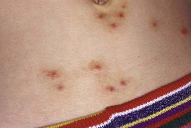
Papular urticaria occurs principally in the first decade of life. It may occur at any time of the year. The most common culprits are species of fleas, mites, bedbugs, gnats, mosquitoes, chiggers, and animal lice. Individuals with papular urticaria have predominantly transitional lesions in various stages of evolution between delayed-onset papules and immediate-onset wheals. The most characteristic lesion is an edematous, red-brown papule (Fig. 660-2). An individual lesion frequently starts as a wheal that, in turn, is replaced by a papule. A given bite may incite an id reaction at distant sites of quiescent bites in the form of erythematous macules, papules, or urticarial plaques. After a season or two, the reaction progresses from a transitional to a primarily immediate hypersensitivity urticarial reaction.
One of the most commonly encountered arthropod bites is that due to human, cat, or dog fleas (family Pulicidae). Eggs, which are generally laid in dusty areas and cracks between floorboards, give rise to larvae that then form cocoons. The cocoon stage can persist for up to 1 yr, and the flea emerges in response to vibrations from footsteps, accounting for the assaults that frequently befall the new owners of a recently reopened dwelling. Adult dog fleas can live without a blood meal for about 60 days. Attacks from fleas are more likely to occur when the fleas do not have access to their usual host; cat or dog fleas are more voracious and problematic when one visits an area frequented by the pet than when the pet is encountered directly. Flea bites tend to be grouped in lines or irregular clusters. Fleas are often not seen on the body of a pet. Diagnosis of flea bites is aided by examination of debris from the animal’s bedding material. The debris is collected by shaking the bedding into a plastic bag and examining the contents for fleas or their eggs, larvae, or feces.


