CHAPTER 1 Anatomy of the brachial plexus
Summary box
Historical perspective
At the end of the 19th century, understanding of the brachial plexus relied on large treatises describing collections of anatomic dissections.1 The strength of these descriptions was fortified by the number of dissections available; however, the quality of these dissections remains uncertain. A number of the anatomic specimens were dissected by medical students, and the accuracy, especially of anatomic variations, is debatable. Many of these descriptions describe copious variations in how the trunks of the plexus coalesce, either into one solid cord, 2 cords, or multiple cords. Many of these early descriptions have not withstood the test of time.
During the same period, a number of works began to codify what was believed to be a “true form” of the plexus. Kerr, Walsh, and Harris2–4 described a series of personally performed or scrutinized dissections that suggested there was far less anatomic variation than previously believed. Convergence of these descriptions of the brachial plexus has allowed a more schematic presentation from which a general foundation can be constructed.
As the understanding of the brachial plexus became more complete, what remained unclear was which cervical roots contributed to it. Some authors believed the plexus was pre-fixed (the plexus originated more cranially than normal and included the C4 nerve root) or that the plexus was post-fixed (the plexus originated more caudally to include the T2 nerve root).1 Other authors believed that instead of having a more cranial or caudal origin, the brachial plexus had a broader or a less broad origin. An even more complex problem was the topographic mapping of nerves within the brachial plexus. A number of authors have pursued microscopic, fascicular dissection of the plexus in order to advance our understanding of the connections within this complex structure.5
Schematic anatomy
The standard schematic diagram used to describe the brachial plexus uses 5 zones: (1) spinal nerve roots, (2) trunks, (3) divisions, (4) cords, and (5) terminal branches.6 The C5 to T1 nerve roots typically contribute to the brachial plexus. The C5 and C6 roots coalesce to form the upper trunk, the C7 root forms the middle trunk, and the C8 and T1 roots coalesce to form the lower trunk. Each trunk divides into an anterior and posterior division. All 3 posterior divisions join to form the posterior cord. The anterior divisions from the upper and middle trunk form the lateral cord, and the anterior division from the lower trunk forms the medial cord. The posterior cord ultimately branches into the terminal branches of the axillary and radial nerves. The lateral cord and medial cord each produce a branch that contributes to form the median nerve. In addition to its contribution to the median nerve, the lateral cord terminates in the musculocutaneous nerve, and the medial cord terminates in the ulnar nerve (Figure 1.1).
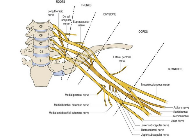
A number of terminal branches (nerves) arise from various zones of the basic structure; knowing these branches and their function facilitates localization of a potential lesion. For instance, the dorsal scapular nerve arises quite proximally from C5, and the long thoracic nerve arises from the nerve roots of C5 to C7; lack of function of either nerve implies a proximal injury of the brachial plexus at the level of the nerve roots. Similarly, the phrenic nerve arises from C3, C4, and C5; diaphragmatic paralysis is also consistent with a proximal lesion of the brachial plexus. The upper trunk gives origin to the suprascapular nerve; lack of supraspinatus and infraspinatus function in the context of deltoid and biceps weakness implies a lesion affecting the upper trunk. More distally, the lateral cord gives rise to the lateral pectoral nerve; the posterior cord gives rise to the upper subscapular nerve, the thoracodorsal nerve, and the lower subscapular nerve; the medial cord gives rise to the medial pectoral nerve, the medial brachial cutaneous nerve, and the medial antebrachial cutaneous nerve (Figure 1.1). Similar logic can be applied to these nerves to determine the site of injury within the brachial plexus.
Surgical anatomy (relationship of the brachial plexus to surrounding structures)
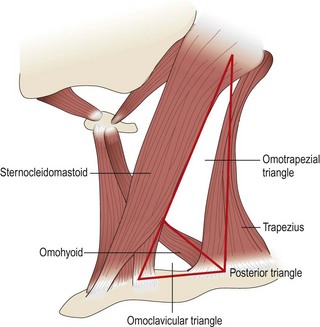
As described above, the brachial plexus has 5 roots (C5-T1), 3 trunks (upper, middle, and lower), 6 divisions (2 divisions, anterior and posterior, per trunk), 3 cords (lateral, posterior, and medial) and 5 main terminal nerve branches (musculocutaneous, radial, axillary, median, and ulnar). Grossly, the brachial plexus emerges in the posterior triangle of the neck (bordered by the sternocleidomastoid and trapezius muscles, clavicle, and occiput). The neck is commonly conceptualized as a set of triangles bounded by identifiable structures. The sternocleidomastoid muscle divides the neck into an anterior and posterior triangle. The omohyoid muscle separates the posterior triangle into a superior, omotrapezial triangle and an inferior, omoclavicular triangle. The upper and middle trunks and their divisions generally lie in the omotrapezial triangle, whereas the lower trunk lies in the omoclavicular triangle (Figure 1.2).
Proximal anatomical relationships
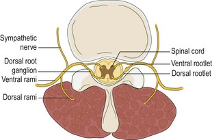
The dorsal rootlet (sensory) and ventral rootlet (motor) converge to form a spinal nerve root. These 2 structures converge approximately at the level of neural foramen (Figure 1.3). The cell bodies of the axons of the sensory rootlet reside in the dorsal root ganglion (outside of the spinal cord), whereas cell bodies of the motor rootlet lie within the anterior horn of the spinal cord. Knowledge of this anatomy not only facilitates intraoperative surgical planning but also the understanding of preoperative electrodiagnostic studies.
< div class='tao-gold-member'>
Stay updated, free articles. Join our Telegram channel

Full access? Get Clinical Tree


