Chapter 515 Anatomic Abnormalities Associated with Hematuria
515.1 Congenital Anomalies
Craig C. Porter and Ellis D. Avner
Gross or microscopic hematuria may be associated with many types of different malformations of the urinary tract. The sudden onset of gross hematuria after minor trauma to the flank is often associated with ureteropelvic junction obstruction or cystic kidneys (see Chapter 531).
515.2 Autosomal Recessive Polycystic Kidney Disease
Also known as infantile polycystic disease, autosomal recessive polycystic kidney disease (ARPKD) is an autosomal recessive disorder occurring with an incidence of 1 : 10,000 to 1 : 40,000. The gene for ARPKD (PKHD1) encodes fibrocystin, a large protein (>4,000 amino acids) with multiple isoforms.
Pathology
Both kidneys are markedly enlarged and grossly show innumerable cysts throughout the cortex and medulla. Microscopic studies demonstrate dilated, ectatic collecting ducts radiating from the medulla to the cortex, although transient proximal tubule cysts have been reported in the fetus. Development of progressive interstitial fibrosis and tubular atrophy during advanced stages of disease eventually leads to renal failure. Liver involvement is characterized by a basic ductal plate abnormality that leads to bile duct proliferation and ectasia, as well as hepatic fibrosis. This lesion is indistinguishable from congenital hepatic fibrosis or Caroli disease, and consequently ARPKD is increasingly referred to as ARPKD/CHF.
Pathogenesis
The function of fibrocystin in normal kidney development and the pathophysiology of its abnormal expression in ARPKD are largely unknown. Evolving information suggests that fibrocystin forms a multimeric complex with proteins of other primary genetic cystic diseases. It appears that altered intracellular signaling from these complexes, located at the apical cell surface, intercellular junction, and basolateral cell surface in association with the focal adhesion complex is a critical feature of disease pathophysiology.
Multiple mutations of PKHD1 cause disease and their average detection rate is approximately 85%. Limited available information suggests some genotype-phenotype correlation: mutations that modify fibrocystin appear to cause less-severe disease than those that truncate fibrocystin.
Clinical Manifestations
The typical child presents with bilateral flank masses during the neonatal period or early infancy. ARPKD may be associated with oligohydramnios, pulmonary hypoplasia, respiratory distress, and spontaneous pneumothorax in the neonatal period. Perinatal demise appears associated with truncating mutations. Components of the oligohydramnios complex including low-set ears, micrognathia, flattened nose, limb-positioning defects, and growth deficiency may be present. Hypertension is usually noted within the first few weeks of life and is often severe and difficult to control. Oliguria and acute renal failure are uncommonly seen, but transient hyponatremia, often in the presence of acute renal failure, often responds to diuresis. Renal function is usually impaired but may be initially normal in 20-30% of patients. Infrequently, ARPKD manifests beyond infancy, in young infants with a mixed clinical picture of renal and hepatic findings: variable degrees of portal hypertension (hepatosplenomegaly, gastroesophageal varices, prominent cutaneous periumbilical veins, reversal of portal vein flow, thrombocytopenia) and variable renal findings that range from asymptomatic abnormal renal ultrasonography to systemic hypertension and renal insufficiency.
In the newborn, clinical evidence of liver disease by radiologic or clinical laboratory assessment is present in about 45% of children. It is believed to be universal by microscopic evaluation. Patients with ARPKD are at risk for developing ascending cholangitis, varices, and hypersplenism related to portal hypertension; they are also at risk for progressive liver fibrosis, which uncommonly leads to overt liver failure and cirrhosis. A subset of older children, and even young adults with ARPKD, present with prominent hepatosplenomegaly and display mild renal disease that is discovered incidentally during imaging studies of the abdomen.
Diagnosis
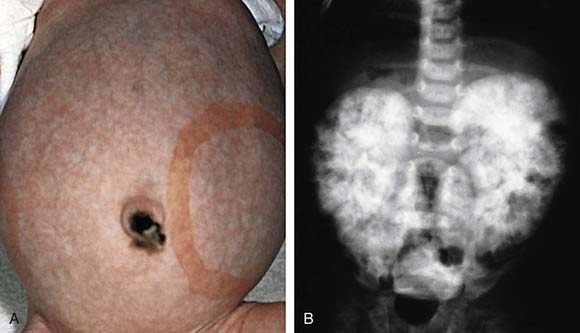
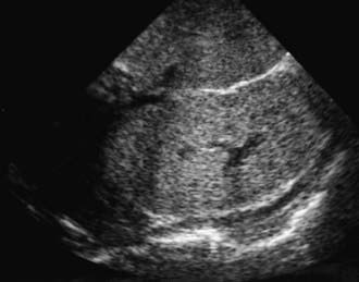
The diagnosis of ARPKD is strongly suggested by bilateral palpable flank masses in an infant with pulmonary hypoplasia, oligohydramnios, and hypertension and the absence of renal cysts by sonography of the parents (Fig. 515-1). Markedly enlarged and uniformly hyperechogenic kidneys with poor corticomedullary differentiation are commonly seen on ultrasonography (Fig. 515-2). The diagnosis is supported by clinical and laboratory signs of hepatic fibrosis, pathologic findings of ductal plate abnormalities seen on liver biopsy, anatomic and pathologic proof of ARPKD in a sibling, or parental consanguinity. The differential diagnosis includes other causes of bilateral renal enlargement and/or cysts, such as multicystic dysplasia, hydronephrosis, Wilms tumor, and bilateral renal vein thrombosis (Table 515-1). Prenatal diagnostic testing using genetic linkage analysis or direct mutation analysis is available in families with ≥1 affected child.

Figure 515-1 A, This infant with infantile polycystic kidney disease shows marked abdominal distention and bilaterally enlarged kidneys, as indicated by the outlined area. B, Intravenous pyelogram of the same patient shows the characteristic mottled nephrogram with brushlike medullary opacification secondary to retention of contrast material in dilated cortical and medullary collecting ducts.
(From Zitelli BJ, Davis HW, editors: Atlas of pediatric physical diagnosis, ed 4, St Louis, 2002, Mosby, p 470.)

Figure 515-2 Ultrasound examination of a neonate with autosomal recessive polycystic kidney disease demonstrating renal enlargement (9 cm) and increased diffuse echogenicity with complete loss of corticomedullary differentiation resulting from multiple small cystic interfaces.

Full access? Get Clinical Tree

