Air-Containing Lesions in Neck
Bernadette L. Koch, MD
DIFFERENTIAL DIAGNOSIS
Common
General Trauma
Retropharyngeal Space (RPS) Abscess
Less Common
Laryngocele
Esophago-Pharyngeal Diverticulum
Rare but Important
4th Branchial Anomaly
Lateral Cervical Esophageal Diverticulum
Spontaneous Cervical Emphysema
ESSENTIAL INFORMATION
Helpful Clues for Common Diagnoses
General Trauma
Key facts: Esophageal, pharyngeal, laryngeal, or superficial trauma
Imaging: Extraluminal air in neck ± laryngeal, hyoid, or facial fractures
Retropharyngeal Space (RPS) Abscess
Key facts: Posterior to pharyngeal mucosal space, anterior to prevertebral space
Imaging: Extranodal purulent fluid in RPS ± air in or adjacent to fluid collection ± extension to mediastinum
Helpful Clues for Less Common Diagnoses
Laryngocele
Key facts: Lateral saccular cyst, laryngeal mucocele
Imaging: Air ± air-filled level
Internal laryngocele in paraglottic space
Mixed laryngocele in paraglottic and submandibular spaces
Esophago-Pharyngeal Diverticulum
Key facts: Zenker diverticulum = mucosal-lined outpouching of posterior hypopharynx
Imaging: Air-filled pouch posterior; usually extends left of esophagus
Helpful Clues for Rare Diagnoses
4th Branchial Anomaly
Key facts: 4th pharyngeal pouch remnant
Extends from apex of pyriform sinus to lower neck, anterior to left thyroid lobe
Present with recurrent thyroiditis or anterior neck abscess
Imaging
Abscess anterior to left thyroid lobe ± intrinsic inflammatory change in ipsilateral thyroid lobe
Inflamed pyriform sinus apex
Lack of aeration of pyriform sinus apex
Lateral Cervical Esophageal Diverticulum
Key facts: Mucosal-lined outpouching lateral to cervical esophagus
Imaging: Air-filled pouch lateral to cervical esophagus
Spontaneous Cervical Emphysema
Key facts: No history of vomiting, trauma, asthma, or other inciting event
Imaging: Pneumomediastinum and cervical emphysema
Image Gallery
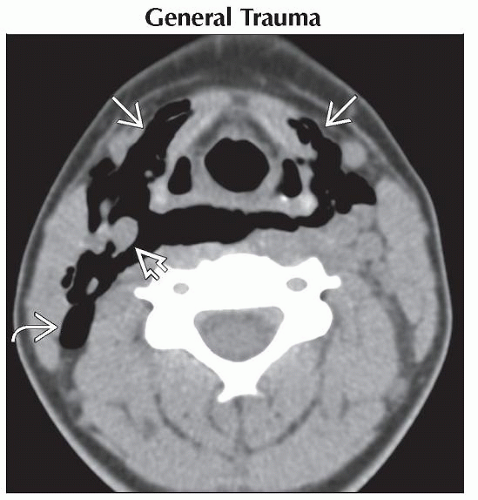 Axial CECT shows extensive air nearly surrounding the larynx
 , surrounding the right carotid vessels , surrounding the right carotid vessels  , and in the posterior triangle of the neck , and in the posterior triangle of the neck  in a child who was thrown from a horse. in a child who was thrown from a horse.Stay updated, free articles. Join our Telegram channel
Full access? Get Clinical Tree
 Get Clinical Tree app for offline access

|