Chapter 421 Acyanotic Congenital Heart Disease
The Obstructive Lesions
421.1 Pulmonary Valve Stenosis with Intact Ventricular Septum
Of the various forms of right ventricular outflow obstruction with an intact ventricular septum, the most common is isolated valvular pulmonary stenosis, which accounts for 7-10% of all congenital heart defects. The valve cusps are deformed to various degrees and, as a result, the valve opens incompletely during systole. The valve may be bicuspid or tricuspid and the leaflets partially fused together with an eccentric outlet. This fusion may be so severe that only a pinhole central opening remains. If the valve is not severely thickened, it produces a dome-like obstruction to right ventricular outflow during systole. Isolated infundibular or subvalvular stenosis, supravalvular pulmonary stenosis, and branch pulmonary artery stenosis are also encountered. In cases where pulmonary valve stenosis is associated with a ventricular septal defect (VSD) but without anterior deviation of the infundibular septum and overriding aorta, this condition is better classified as pulmonary stenosis with VSD rather than as tetralogy of Fallot (Chapter 424.1). Pulmonary stenosis and an atrial septal defect (ASD) are also occasionally seen as associated defects. The clinical and laboratory findings reflect the dominant lesion, but it is important to rule out any associated anomalies. Pulmonary stenosis as a result of valve dysplasia is the most common cardiac abnormality in Noonan syndrome (Chapter 76), and is associated in about 50% of cases with a mutation in the gene PTPN11, encoding the protein tyrosine phosphotase SHP-2 on chromosome 12. The mechanism for pulmonic stenosis is unknown, although maldevelopment of the distal portion of the bulbus cordis and the sequelae of fetal endocarditis have been suggested as etiologies. Pulmonary stenosis, either of the valve or the branch pulmonary arteries, is a common finding in patients with arteriohepatic dysplasia, also known as Alagille syndrome (Chapter 348). In this syndrome and in some patients with isolated pulmonic stenosis, a mutation is present in the Jagged1 gene.
Pathophysiology
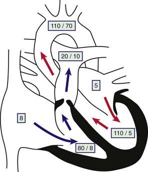
The obstruction to outflow from the right ventricle to the pulmonary artery results in increased right ventricular systolic pressure and wall stress, which leads to hypertrophy of the right ventricle (Fig. 421-1). The severity of these abnormalities depends on the size of the restricted valve opening. In severe cases, right ventricular pressure may be higher than systemic arterial systolic pressure, whereas with milder obstruction, right ventricular pressure is only mildly or moderately elevated. Pulmonary artery pressure (distal to the obstruction) is normal or decreased. Arterial oxygen saturation will be normal even in cases of severe stenosis, unless an intracardiac communication such as a VSD or ASD is allowing blood to shunt from right to left. When severe pulmonic stenosis occurs in a neonate, decreased right ventricular compliance often leads to cyanosis due to right-to-left shunting through a patent foramen ovale, a condition termed critical pulmonic stenosis.

Figure 421-1 Physiology of valvular pulmonary stenosis. Boxed numbers represent pressure in mm Hg. Because of the absence of right-to-left or left-to-right shunting, blood flow through all cardiac chambers is normal at 3 L/min/m2. The pulmonary-to-systemic blood flow ratio (Qp : Qs) is 1 : 1. Right atrial pressure is increased slightly as a result of decreased right ventricular compliance. The right ventricle is hypertrophied, and systolic and diastolic pressure is increased. The pressure gradient across the thickened pulmonary valve is 60 mm Hg. The main pulmonary artery pressure is slightly low, and poststenotic dilatation is present. Left heart pressure is normal. Unless right-to-left shunting is occurring through a foramen ovale, the patient’s systemic oxygen saturation will be normal.
Clinical Manifestations and Laboratory Findings
Patients with mild or moderate stenosis usually do not have any symptoms. Growth and development are most often normal. If the stenosis is severe, signs of right ventricular failure such as hepatomegaly, peripheral edema, and exercise intolerance may be present. In a neonate or young infant with critical pulmonic stenosis, signs of right ventricular failure may be more prominent, and cyanosis is often present because of right-to-left shunting at the foramen ovale.
With mild pulmonary stenosis, venous pressure and pulse are normal. The heart is not enlarged, the apical impulse is normal, and the right ventricular impulse is not palpable. A sharp pulmonic ejection click immediately after the 1st heart sound is heard at the left upper sternal border during expiration. The 2nd heart sound is split, with a pulmonary component of normal intensity that may be slightly delayed. A relatively short, low- or medium-pitched systolic ejection murmur is maximally audible over the pulmonic area and radiates minimally to the lung fields bilaterally. The electrocardiogram is normal or characteristic of mild right ventricular hypertrophy; inversion of the T waves in the right precordial leads may be seen. (Remember that the T wave in lead V1 should normally be inverted until at least 6-8 yr of age. Therefore, a positive T wave in V1 in a young child is a sign of right ventricular hypertrophy.) The only abnormality demonstrable radiographically is usually poststenotic dilatation of the pulmonary artery. Two-dimensional echocardiography shows right ventricular hypertrophy and a slightly thickened pulmonic valve, which domes in systole; Doppler studies demonstrate a right ventricle to pulmonary artery gradient of ≤30 mm Hg.
In moderate pulmonic stenosis, venous pressure may be slightly elevated; in older children, a prominent a wave may be noted in the jugular pulse. A right ventricular lift may be palpable at the lower left sternal border. The 2nd heart sound is split, with a delayed and soft pulmonary component. As valve motion becomes more limited with more severe degrees of stenosis, both the pulmonic ejection click and the pulmonic 2nd sound may become inaudible. With increasing degrees of stenosis, the peak of the systolic ejection murmur is prolonged later into systole, and its quality becomes louder and harsher (higher frequency). The murmur radiates more prominently to both lung fields.
The electrocardiogram reveals right ventricular hypertrophy, sometimes with a prominent spiked P wave. Radiographically, the heart can vary from normal size to mildly enlarged with uptilting of the apex due to prominence of the right ventricle; pulmonary vascularity may be normal or slightly decreased. The echocardiogram shows a thickened pulmonic valve with restricted systolic motion. Doppler examination demonstrates a right ventricle to pulmonary artery pressure gradient in the 30-60 mm Hg range. Mild tricuspid regurgitation may be present and allows Doppler confirmation of right ventricular systolic pressure.
In severe stenosis, mild to moderate cyanosis may be noted in patients with an interatrial communication (atrial septal defect or patent foramen ovale). If hepatic enlargement and peripheral edema are present, they are an indication of right ventricular failure. Elevation of venous pressure is common and is caused by a large presystolic jugular a wave. The heart is moderately or greatly enlarged, and a conspicuous parasternal right ventricular lift is present and frequently extends to the left midclavicular line. The pulmonary component of the 2nd sound is usually inaudible. A loud, long, and harsh systolic ejection murmur, usually accompanied by a thrill, is maximally audible in the pulmonic area and may radiate over the entire precordium, to both lung fields, into the neck, and to the back. The peak of the murmur occurs later in systole as valve opening becomes more restricted. The murmur frequently encompasses the aortic component of the 2nd sound but is not preceded by an ejection click.
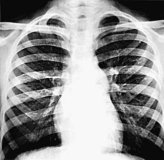
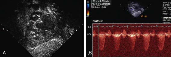
The electrocardiogram shows gross right ventricular hypertrophy, frequently accompanied by a tall, spiked P wave. Radiographic studies confirm the presence of cardiac enlargement with prominence of the right ventricle and right atrium. Prominence of the main pulmonary artery segment may be seen due to poststenotic dilatation (Fig. 421-2). Intrapulmonary vascularity is decreased. The two-dimensional echocardiogram shows severe deformity of the pulmonary valve and right ventricular hypertrophy (Fig. 421-3). In the late stages of the disease, systolic dysfunction of the right ventricle may be seen, and in these cases the ventricle may become dilated, with prominent tricuspid regurgitation. Doppler studies demonstrate a high gradient (>60 mm Hg) across the pulmonary valve. The classic findings of severe pulmonary stenosis in older children are rarely seen because of early intervention. Signs of critical pulmonic stenosis, with all of the features of severe pulmonic stenosis plus cyanosis, are usually encountered in the neonatal period.

Figure 421-2 Roentgenogram in a patient with valvular pulmonary stenosis and a normal aortic root. The heart size is within normal limits, but poststenotic dilatation of the pulmonary artery is present.

Figure 421-3 Echocardiogram demonstrating valvar pulmonic stenosis. A, Subcostal view showing thickened pulmonary valve leaflets (between crosshatches). B, Doppler study indicating a 95 mm Hg peak pressure gradient across the stenotic valve. MPA, main pulmonary artery; RV, right ventricle.
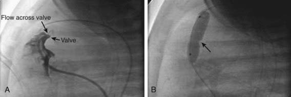
Cardiac catheterization is not generally required for diagnostic purposes but is undertaken as part of a balloon valvuloplasty procedure. Catheterization demonstrates an abrupt pressure gradient across the pulmonary valve. Pulmonary artery pressure is either normal or low. The severity of the stenosis is graded based on the ratio of right ventricular systolic pressure to systemic systolic pressure or the right ventricle to pulmonary artery pressure gradient: a gradient of 10-30 mm Hg in mild cases, 30-60 mm Hg in moderate cases, and >60 mm Hg or with right ventricular pressure greater than systemic pressure in severe cases. If cardiac output is low or a significant right-to-left shunt exists across the atrial septum, the pressure gradient may underestimate the degree of valve stenosis. Selective right ventriculography demonstrates the thickened, poorly mobile valve. In mild to moderate stenosis, doming of the valve in systole is readily seen. Flow of contrast medium through the stenotic valve in ventricular systole produces a narrow jet of dye that fills the dilated main pulmonary artery. Subvalvular hypertrophy that may intensify the obstruction may be present.
Treatment
Patients with moderate or severe isolated pulmonary stenosis require relief of the obstruction. Balloon valvuloplasty is the initial treatment of choice for the majority of patients (Fig. 421-4). Patients with severely thickened pulmonic valves, especially common in those with Noonan syndrome, may require surgical intervention. In a neonate with critical pulmonic stenosis, urgent treatment by either balloon valvuloplasty or surgical valvotomy is warranted.

Figure 421-4 Valvar pulmonary stenosis and balloon valvuloplasty. A, Right ventricular angiogram showing severely stenotic pulmonary valve with narrow jet of blood flowing across. B, Inflation of the balloon catheter showing the indentation (arrow) made on the balloon from the stenotic valve.
(Photos courtesy of Dr. Jeffrey Feinstein, Stanford University, Stanford, CA.)
Excellent results are obtained in most instances. The gradient across the pulmonary valve is markedly reduced or abolished. In the early period after balloon valvuloplasty, a small to moderate residual gradient may remain because of muscular infundibular narrowing; it usually resolves with time. A short, early decrescendo diastolic murmur may be heard at the mid to upper left sternal border as a result of pulmonary valvular insufficiency. The degree of insufficiency is not usually clinically significant. No difference in patient status after valvuloplasty or surgery is noted at late follow-up; recurrence is unusual after successful treatment except in those patients with extremely dysplastic valves.
Prognosis and Complications
Heart failure occurs only in severe cases and most often during the 1st mo of life. The development of cyanosis from a right-to-left shunt across a foramen ovale is almost exclusively seen in the neonatal period when the stenosis is severe. Infective endocarditis is a risk but is not common in childhood.
Children with mild stenosis can lead a normal life, but their progress should be evaluated at regular intervals. Patients who have small gradients rarely show progression and do not need intervention, but a significant gradient is more likely to develop in children with moderate stenosis as they grow older. Worsening of obstruction may also be due to the development of secondary subvalvular muscular and fibrous tissue hypertrophy. In untreated severe stenosis, the course may abruptly worsen with the development of right ventricular dysfunction and cardiac failure. Infants with critical pulmonic stenosis require urgent catheter balloon valvuloplasty or surgical valvotomy. Development of right ventricular failure many years after pulmonary balloon valvuloplasty is uncommon. Nonetheless, patients should be followed serially for worsening pulmonary insufficiency and right ventricular dilation.
Crosnier C, Lykavieris P, Meunier-Rotival M, et al. Alagille syndrome. The widening spectrum of arteriohepatic dysplasia. Clin Liver Dis. 2000;4:765-778.
Feinstein JA, Kim N, Reddy VM, Perry SB. Percutaneous pulmonary valve placement in a 10-month-old patient using a hand crafted stent-mounted porcine valve. Catheter Cardiovasc Interv. 2006;67:644-649.
Khambadkone S, Coats L, Taylor A, et al. Percutaneous pulmonary valve implantation in humans: results in 59 consecutive patients. Circulation. 2005;112:1189-1197.
Krantz ID, Smith R, Colliton RP, et al. Jagged 1 mutations in patients ascertained with isolated congenital heart defects. Am J Med Genet. 1999;84:56-60.
Phoon CK. Estimation of pressure gradients by auscultation: an innovative and accurate physical examination technique. Am Heart J. 2001;141:500-506.
Rosales AM, Lock JE, Perry SB, et al. Interventional catheterization management of perioperative peripheral pulmonary stenosis: balloon angioplasty or endovascular stenting. Catheter Cardiovasc Interv. 2002;56:272-277.
Yoshida R, Hasegawa T, Hasegawa Y, et al. Protein-tyrosine phosphatase, nonreceptor type 11 mutation analysis and clinical assessment in 45 patients with Noonan syndrome. J Clin Endocrinol Metab. 2004;89:3359-3364.
421.2 Infundibular Pulmonary Stenosis and Double-Chamber Right Ventricle
Infundibular pulmonary stenosis is caused by muscular or fibrous obstruction in the outflow tract of the right ventricle. The site of obstruction may be close to the pulmonary valve or well below it; an infundibular chamber may be present between the right ventricular cavity and the pulmonary valve. In many cases, a VSD may have been present initially and later closed spontaneously. When the pulmonary valve is also stenotic, the combined defect is primarily classified as valvular stenosis with secondary infundibular hypertrophy. The hemodynamics and clinical manifestations of patients with isolated infundibular pulmonary stenosis are similar, for the most part, to those described in the discussion of isolated valvular pulmonary stenosis (Chapter 421.1).
A common variation in right ventricular outflow obstruction below the pulmonary valve is that of a double-chambered right ventricle. In this condition, a muscular band is present in the mid-right ventricular region; the band divides the chamber into two parts and creates obstruction between the inlet and outlet portions. An associated VSD that may close spontaneously is often noted. Obstruction is not usually seen early in life but may progress rapidly in a similar manner to the progressive infundibular obstruction observed with tetralogy of Fallot (Chapter 424.1).
421.3 Pulmonary Stenosis in Combination with an Intracardiac Shunt
Valvular or infundibular pulmonary stenosis, or both, may be associated with either an ASD or a VSD. In these patients, the clinical features depend on the degree of pulmonary stenosis, which determines whether the net shunt is from left to right or from right to left.
The presence of a large left-to-right shunt at the atrial or ventricular level is evidence that the pulmonary stenosis is mild. These patients have symptoms similar to those of patients with an isolated ASD or VSD. With increasing age, worsening of the obstruction may limit the shunt and result in a gradual improvement in symptoms. Eventually, particularly in patients with pulmonary stenosis and VSD, a further increase in obstruction may lead to right-to-left shunting and cyanosis. When a patient with a VSD has evidence of decreasing heart failure and increased right ventricular forces on the electrocardiogram, one must differentiate between the development of increasing pulmonary stenosis versus the onset of pulmonary vascular disease (Eisenmenger syndrome, Chapter 427.2).
These anomalies are readily repaired surgically. Defects in the atrial or ventricular septum are closed, and the pulmonary stenosis is relieved by resection of infundibular muscle or pulmonary valvotomy, or both, as indicated. Patients with a predominant right-to-left shunt have symptoms similar to those of patients with tetralogy of Fallot (Chapter 424.1).
421.4 Peripheral Pulmonary Stenosis
Single or multiple constrictions may occur anywhere along the major branches of the pulmonary arteries and may range from mild to severe and from localized to extensive. Frequently, these defects are associated with other types of congenital heart disease, including valvular pulmonic stenosis, tetralogy of Fallot, patent ductus arteriosus (PDA), VSD, ASD, and supravalvular aortic stenosis. A familial tendency has been recognized in some patients with peripheral pulmonic stenosis. A high incidence is found in infants with congenital rubella syndrome. The combination of supravalvular aortic stenosis with pulmonary arterial branch stenosis, idiopathic hypercalcemia of infancy, elfin facies, and mental retardation is known as Williams syndrome, a condition associated with deletion of the elastin gene in region 7q11.23 on chromosome 7. Peripheral pulmonary stenosis is also associated with the Alagille syndrome, which may be associated with a mutation in the Jagged1 gene.
A mild constriction has little effect on the pulmonary circulation. With multiple severe constrictions, pressure is increased in the right ventricle and in the pulmonary artery proximal to the site of obstruction. When the anomaly is isolated, the diagnosis is suspected by the presence of murmurs in widespread locations over the chest, either anteriorly or posteriorly. These murmurs are usually systolic ejection in quality but may be continuous. Most often, the physical signs are dominated by the associated anomaly, such as tetralogy of Fallot (Chapter 424.1).
If the stenosis is severe, the electrocardiogram shows evidence of right ventricular and right atrial hypertrophy, and the chest radiograph shows cardiomegaly and prominence of the main pulmonary artery. The pulmonary vasculature is usually normal; in some cases, however, small intrapulmonary vascular shadows are seen that represent areas of poststenotic dilatation. Echocardiography is limited in its ability to visualize the distal branch pulmonary arteries. Doppler examination demonstrates the acceleration of blood flow through the stenoses and, if tricuspid regurgitation is present, allows an estimation of right ventricular systolic pressure. MRI and CT are extremely helpful in delineating distal obstructions; if moderate to severe disease is suspected, the diagnosis is usually confirmed by cardiac catheterization.
Severe obstruction of the main pulmonary artery and its primary branches can be relieved during corrective surgery for associated lesions such as the tetralogy of Fallot or valvular pulmonary stenosis. If peripheral pulmonic stenosis is isolated, it may be treated by catheter balloon dilatation, sometimes with placement of an intravascular stent (Fig. 417-29).
421.5 Aortic Stenosis
Pathophysiology
Congenital aortic stenosis accounts for ≈5% of cardiac malformations recognized in childhood; a bicuspid aortic valve, one of the most common congenital heart lesions overall, is identified in up to 1.5% of adults and may be asymptomatic in childhood. Aortic stenosis is more frequent in males (3 : 1). In the most common form, valvular aortic stenosis, the leaflets are thickened and the commissures are fused to varying degrees. Left ventricular systolic pressure is increased as a result of the obstruction to outflow. The left ventricular wall hypertrophies in compensation; as its compliance decreases, end-diastolic pressure increases as well.
Subvalvular (subaortic) stenosis with a discrete fibromuscular shelf below the aortic valve is also an important form of left ventricular outflow tract obstruction. This lesion is frequently associated with other forms of congenital heart disease such as mitral stenosis and coarctation of the aorta (Shone syndrome

Stay updated, free articles. Join our Telegram channel

Full access? Get Clinical Tree