Absent Kidney
Paula J. Woodward, MD
DIFFERENTIAL DIAGNOSIS
Common
Renal Agenesis
Bilateral Renal Agenesis
Unilateral Renal Agenesis
Less Common
Mimics for Renal Agenesis
Pelvic Kidney
Crossed Fused Ectopia
ESSENTIAL INFORMATION
Key Differential Diagnosis Issues
Is the kidney truly absent?
Always search for ectopic location
Is the fluid normal?
Bilateral renal agenesis will have anhydramnios
Remainder have normal fluid
Fetal adrenal is large and easily mistaken for a kidney, especially in 1st and 2nd trimester
Normal adrenal has an “ice cream sandwich” appearance
Hypoechoic cortex surrounding a hyperechoic medulla
In renal agenesis adrenal has a flattened, discoid, “lying down” appearance
Adrenal gland does not fold into “Y” or “tricorn hat” configuration if no kidney
Helpful Clues for Common Diagnoses
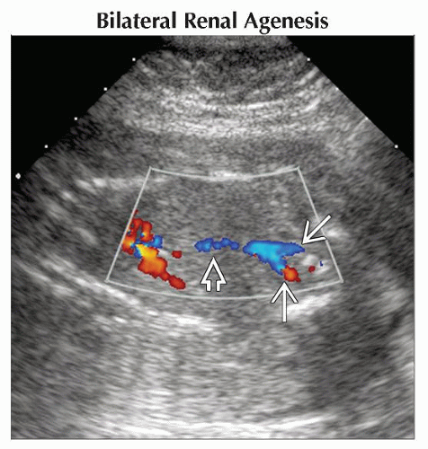
Bilateral Renal Agenesis
No demonstrable renal tissue
No urine in fetal bladder
Anhydramnios
“Lying down” adrenals in renal fossa, although this may be difficult to see in setting of no fluid
Look for renal arteries, but be aware of pitfalls
Lumbar arteries can easily be mistaken for renal arteries
MR very helpful for confirmation of diagnosis
Unilateral Renal Agenesis
One kidney seen, which may show compensatory hypertrophy
May see “lying down” adrenal ipsilateral to absent kidney
Bladder seen to fill and empty
Normal amniotic fluid volume
Helpful Clues for Less Common Diagnoses
Pelvic Kidney
Empty renal fossa
Kidney in fetal pelvis, superior to bladder
May be difficult to see as echogenicity similar to bowel
Contralateral kidney is normal-sized
Crossed Fused Ectopia
Ectopic kidney located in opposite flank creating a large bilobed kidney
95% fused, 5% unfused
Left crosses to right most often
Other Essential Information
Uterine anomalies associated with renal anomalies, especially renal agenesis
Image Gallery
 Coronal color Doppler ultrasound of a fetus with anhydramnios, which makes visualization of fetal anatomy difficult. Flow is seen in the aorta
 and iliac arteries and iliac arteries  , but no renal arteries were identified. , but no renal arteries were identified.Stay updated, free articles. Join our Telegram channel
Full access? Get Clinical Tree
 Get Clinical Tree app for offline access

|