Chapter 623 Abnormalities of the Optic Nerve
Optic Nerve Aplasia
Optic nerve aplasia is a rare congenital anomaly and is typically unilateral. The optic nerve, retinal ganglion cells, and retinal blood vessels are absent. A vestigial dural sheath usually connects with the sclera in a normal position, but no neural tissue is present within this sheath. Optic nerve aplasia typically occurs sporadically in an otherwise healthy person. A wide variety of ocular abnormalities can occur, but colobomas are the most common associated finding.
Optic Nerve Hypoplasia
Hypoplasia of the optic nerve is a nonprogressive condition characterized by a subnormal number of optic nerve axons with normal mesodermal elements and glial supporting tissue. In typical cases, the nerve head is small and pale, with a pale or pigmented peripapillary halo or double-ring sign.
This anomaly is associated with defects of vision and of visual fields of varying severity, ranging from blindness to normal or near-normal vision. It may be associated with systemic anomalies that most commonly involve the central nervous system (CNS). Protean CNS defects such as hydranencephaly or anencephaly or more focal lesions compatible with continued development of a patient can accompany optic nerve hypoplasia, but unilateral or bilateral optic nerve hypoplasia may be found without any concomitant defects.
Optic nerve hypoplasia is a principal feature of septo-optic dysplasia of de Morsier, a developmental disorder characterized by the association of anomalies of the midline structures of the brain with hypoplasia of the optic nerves, optic chiasm, and optic tracts; typically noted are agenesis of the septum pellucidum, partial or complete agenesis of the corpus callosum, and malformation of the fornix, with a large chiasmatic cistern. Patients can have hypothalamic abnormalities and endocrine defects ranging from panhypopituitarism to isolated deficiency of growth hormone, hypothyroidism, or diabetes insipidus. Neonatal hypoglycemia and seizures are important presenting signs in affected infants (Chapter 585).
Bilateral subtle hypoplasia may be difficult to diagnose from the appearance of the disc alone because no comparison with a contralateral uninvolved eye is possible. However, it is important to establish the diagnosis because this eliminates confusion with optic atrophy or glaucoma and can explain the cause of decreased vision in a patient unresponsive to amblyopia therapy. Endocrine function should be watched closely in patients with optic nerve hypoplasia.
The cause of optic nerve hypoplasia remains unclear. Early gestational injuries to midline CNS structures with secondary axonal injury or a disruption of normal neuronal guidance mechanisms that affect both the optic nerve and cerebral neurons might account for these commonly associated disorders. Optic nerve hypoplasia can occur with somewhat increased incidence in infants of diabetic mothers and has been associated with maternal use of phenytoin, quinine, LSD (lysergic acid diethylamide), and alcohol during pregnancy. Preterm labor, gestational vaginal bleeding, low maternal weight gain, and weight loss during pregnancy in young primaparas have also been identified as risk factors.
Children with periventricular leukomalacia display an unusual form of optic nerve hypoplasia. The optic nerves demonstrate a large cup within a normal-sized optic disc. This form of optic nerve hypoplasia occurs secondary to transsynaptic degeneration of optic axons caused by the primary bilateral lesion in the optic radiation (periventricular leukomalacia).
Optic Nerve Coloboma
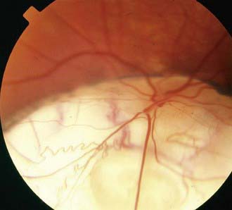
Optic nerve colobomas can be unilateral or bilateral. The visual acuity can range from normal to complete blindness. The coloboma develops secondary to incomplete closure of the embryonic fissure. The defect can produce a partial or total excavation of the optic disc (Fig. 623-1) Chorioretinal and iris colobomas can also occur. Optic nerve colobomas may be seen in a multitude of ocular and systemic abnormalities including the CHARGE association (coloboma, heart disease, atresia choanae, retarded growth and development and/or central nervous system anomalies, genetic anomalies and/or hypogonadism, ear anomalies and/or deafness).
Morning Glory Disc Anomaly
Morning glory disc anomaly is a congenital malformation of the optic nerve characterized by an enlarged, excavated, funnel-shaped disc with an elevated rim, resembling a morning glory flower. White glial tissue is present in the central part of the disc. The retinal vessels are abnormal and appear at the peripheral disc and course over the elevated pink rim in a radial fashion. Pigmentary mottling of the peripapillary region is usually seen. Most cases are unilateral. Girls are affected twice as often as boys. Visual acuity is usually severely reduced, and retinal detachment occurs in approximately of involved eyes. Morning glory disc anomaly has been associated with basal encephalocele in patients with midfacial anomalies. Abnormalities of the carotid circulation can also be seen in patients with morning glory anomaly. Moyamoya disease is a well-described associated finding.
Tilted Disc
Tilted disc is a congenital anomaly in which the vertical axis of the optic disc is directed obliquely, so that the upper temporal portion of the nerve head is more prominent and anterior to the lower nasal portion of the disc. The retinal vessels emerge from the upper temporal portion of the disc rather than from the nasal side. Often noted is a peripapillary crescent or conus. Associated visual field defects and myopic astigmatism may be found. Clinical recognition of the tilted disc syndrome is important to avoid confusion of its disc and visual field signs with those of papilledema and intracranial tumor.

Full access? Get Clinical Tree

