Chapter 614 Abnormalities of Pupil and Iris
Aniridia
The term aniridia is a misnomer because iris tissue is usually present, although it is hypoplastic (Fig. 614-1). Two thirds of the cases are dominantly transmitted with a high degree of penetrance. The other 30% of cases are sporadic and are considered to be new mutations. The condition is bilateral in 98% of all patients, regardless of the means of transmission, and is found in approximately 1/50,000 persons.

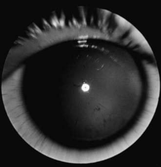
Figure 614-1 Aniridia with minimal iris tissue.
(From Nelson LB, Spaeth GL, Nowinski TS, et al: Aniridia: a review, Surv Ophthalmol 28:621–642, 1984.)
Aniridia is a panocular disorder and should not be thought of as an isolated iris defect. Macular and optic nerve hypoplasias are commonly present and lead to decreased vision and sensory nystagmus. The visual acuity is measured as 20/200 in most patients, although the vision occasionally is better. Other ocular deformities are common and can involve the lens and cornea. The cornea may be small, and a cellular infiltrate (pannus) occasionally develops in the superficial layers of the peripheral cornea. Clinically, this appears as a gray opacification. The pannus results from a stem cell deficiency and therefore must be treated with keratolimbal stem cell transplantation rather than cornea transplantation. Lens abnormalities include cataract formation and partial or total lens dislocation. Glaucoma develops in as many as 75% of patients with aniridia.
Aniridia is caused by a defect in the PAX6 gene on chromosome 11p13. The PAX6 gene is the master control gene for eye morphogenesis. Aniridia can be sporadic or familial. The familial form is autosomal dominant with complete penetrance but variable expressivity. Sporadic aniridia is associated with Wilms tumor in as many as 30% of cases (Chapter 493.1). The combination of aniridia and Wilms tumor represents a contiguous gene syndrome in which the adjacent PAX6 and Wilms tumor (WT1) genes are both deleted. Some deletions create the WAGR complex of Wilms tumor, aniridia, genitourinary malformations, and mental retardation. All children with sporadic aniridia should undergo chromosomal deletional analysis to exclude the possibility of Wilms tumor formation. Children who test positive for the deletion should undergo repeated abdominal ultrasonographic and clinical examinations. Wilms tumor has also been reported in patients with familial aniridia. Therefore, these patients should also undergo chromosomal analysis.
Coloboma of the Iris
Coloboma of the iris is a developmental defect that can occur as a defect in a sector of the iris, a hole in the substance of the iris, or a notch in the pupil’s margin. Simple colobomas are often transmitted as an autosomal dominant trait and can occur alone or in association with other anomalies. A coloboma is formed when the embryonic fissure fails to close completely. Because of the anatomic location of the embryonic fissure, an iris coloboma is always located inferiorly, giving the iris a keyhole appearance. An iris coloboma may be the only externally visible part of an extensive malclosure of the embryonic fissure that also involves the fundus and optic nerve. When this occurs, vision is likely to be severely affected. Therefore, all children with an iris coloboma should undergo a full ophthalmologic examination.
Microcoria
Microcoria (congenital miosis) appears as a small pupil that does not react to light or accommodation and that dilates poorly, if at all, with medication. The condition may be unilateral or bilateral. In bilateral cases, the degree of miosis may be different in each eye. The eye may be otherwise normal or may demonstrate other abnormalities of the anterior segment. Congenital microcoria is usually transmitted as an autosomal dominant trait, although it can occur sporadically.
Congenital Mydriasis
In congenital mydriasis, the pupils appear dilated, do not constrict significantly to light or near gaze, and respond minimally to miotic agents. The iris is otherwise normal, and affected children are usually healthy. Trauma, pharmacologic mydriasis, and neurologic disorders should be considered. Many apparent cases of congenital mydriasis show abnormalities of the central iris structures and may be considered a form of aniridia.
Dyscoria and Corectopia
Dyscoria is abnormal shape of the pupil, and corectopia is abnormal position of the pupil. They can occur together or independently as congenital or acquired anomalies.
Congenital corectopia is usually bilateral and symmetric and rarely occurs as an isolated anomaly; it is usually accompanied by dislocation of the lens (ectopia lentis et pupillae), and the lens and pupil are commonly dislocated in opposite directions. Ectopia lentis et pupillae is transmitted as an autosomal recessive disorder; consanguinity is common.
When these are acquired, distortion and displacement of the pupil are often a result of trauma or intraocular inflammation. Prolapse of the iris after perforating injuries of the eye leads to peaking of the pupil in the direction of the perforation. Posterior synechiae (adhesions of the iris to the lens) are commonly seen when inflammation due to any cause occurs in the anterior segment.
Anisocoria
This is inequality of the pupils. The difference in size may be due to local or neurologic disorders. As a rule, if the inequality is more pronounced in the presence of bright focal illumination or on near gaze, there is a defect in pupillary constriction and the larger pupil is abnormal. If the anisocoria is worse in reduced illumination, a defect in dilation exists and the smaller pupil is abnormal. Neurologic causes of anisocoria (parasympathetic or sympathetic lesions) must be differentiated from local causes such as synechiae (adhesions), congenital iris defects (colobomas, aniridia), and pharmacologic effects. Simple central anisocoria can occur in otherwise healthy children. The combination of anisocoria and ptosis may be seen in Horner syndrome.

Stay updated, free articles. Join our Telegram channel

Full access? Get Clinical Tree

