Heterozygous mutations in the TCF2 (transcription factor 2) gene are an important cause of bilateral hyperechogenic kidneys and kidneys with cystic dysplasia. The phenotype associated with TCF2 gene mutations is highly variable, even among members of the same family. Extrarenal findings may be present and include urogenital abnormalities and maturity-onset diabetes of the young.
Autosomal dominant polycystic kidney disease (PKD) is one of the most common multisystem genetic disorders among adults with an incidence of about 1 in 800. Almost all affected individuals have a mutation in one of two genes, PKD1 or PKD2, which account for 85% and 15% of cases, respectively. Clinical onset of kidney disease is usually between 30 and 50 years but can vary widely among affected individuals in the same family. Cases with prenatal onset of renal findings identifiable by ultrasonographic examination occur rarely. Prenatal findings include enlarged kidneys, hyperechogenic renal cortex and medulla, and diminished or absent corticomedullary differentiation. Small bilateral cortical cysts may or may not be present. Amniotic fluid volume is normal. In rare families, unilateral renal hypoplasia or aplasia has been reported.
Occasionally the diagnosis of autosomal dominant PKD is first made in a fetus or in a young child who has a presymptomatic parent. Also, about 5% of cases are due to de novo mutations in PKD1 or PKD2 genes. In the absence of a family history of the disorder, autosomal dominant polycystic kidney disease has only a small chance of being the cause of echogenic cystic kidneys.
Autosomal recessive polycystic kidney disease (infantile polycystic kidney disease) is a rare disorder occurring in 1 in 20 000 to 1 in 50 000 births. There is a wide range of interfamilial and intrafamilial phenotypic severity and age of onset. Fifty percent of cases are identifiable by prenatal ultrasonography. Kidneys can be grossly enlarged resulting in a large abdominal circumference with echogenic renal parenchyma and loss of corticomedullary differentiation. However, renal cysts are not evident in the first or second trimester in most prenatally diagnosed cases. Amniotic fluid volume ranges from normal to absent.
Mutations in one gene, PKHD1 (polycystic kidney and hepatic disease 1), cause autosomal recessive polycystic kidney disease. There is no evidence for a second locus.
In order to maximize the chance of a establishing a diagnosis for the fetus, further prenatal testing should include documentation of the presence or absence of extrarenal abnormalities by prenatal ultrasonographic examination and renal ultrasonographic examinations for both parents. Fetal chromosome analysis can be obtained by chorionic villus sampling if severe oligohydramnios is present, or by bladder puncture for urinary tract cells if the bladder is distended. After delivery, the baby should have a careful examination by a nephrologist. If the baby does not survive, a complete pathologic examination of the fetus is indicated. In addition, DNA should be obtained from fetal tissue for possible molecular diagnostic testing in the future.
Further Reading
1. Aubertin G, Cripps S, Coleman G et al. (2002) Prenatal diagnosis of apparently isolated unilateral multicystic kidney: implications for counselling and management. Prenatal Diagnosis 22:388–394.
2. Bisceglia M, Galliani CA, Senger C et al. (2006) Renal cystic diseases: a review. Advances in Anatomic Pathology 13:26–56.
3. Decramer S, Parant O, Beaufils S et al. (2007) Anomalies of the TCF2 gene are the main cause of fetal bilateral hyperechogenic kidneys. Journal of the American Society of Nephrology 18:923–933.
4. Harris P, Rossetti S (2004) Molecular genetics of autosomal recessive polycystic kidney disease. Molecular Genetics and Metabolism 81:75–85.
5. Pei Y (2003) Molecular genetics of autosomal dominant polycystic kidney disease. Clinical and Investigative Medicine 26:252–258.
6. Winyard P, Chitty L (2001) Dysplastic and polycystic kidneys: diagnosis, associations and management. Prenatal Diagnosis 21:924–935.
Omphalocele
Ultrasonographic examination performed at 17 weeks in a 37-year-old woman due to an elevated serum AFP concentration revealed a fetal omphalocele. Fetal growth and all other fetal anatomy appeared normal. Amniocentesis revealed a normal female karyotype. Fetal echocardiography at 22 weeks’ gestation was also normal. The pregnancy was conceived via in vitro fertilization due to several years of unexplained infertility. Both parents have normal peripheral blood karyotypes and the husband has normal sperm parameters. First trimester screening predicted low risks for Down syndrome and trisomies 18 and 13.
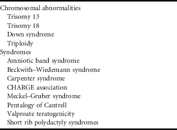
Omphalocele can occur as an isolated abnormality or as one feature of a number of Mendelian and chromosomal disorders (Table 5.2).
Table 5.2 Disorders in which omphalocele is a feature
Reproduced from Sanders RC (2002) Structural Fetal Abnormalities, 2nd edition. Mosby, St Louis MO; London reproduced with permission from Elsevier.
In about two-thirds of cases of omphalocele, there are associated malformations including heart defects, neural tube defects, facial clefting, diaphragmatic hernia, bladder extrophy, and imperforate anus. With the exception of imperforate anus, most of these abnormalities should be evident on prenatal ultrasonographic examination.
When omphalocele is the only finding on ultrasonographic examination and the fetal karyotype is normal, there still remains a several percent risk of a genetic syndrome although an isolated omphalocele remains the most likely outcome. Of the possible genetic syndromes which could be present, Beckwith–Wiedemann syndrome (BWS) would be the most likely with an estimated chance of 5–20%.
BWS is a generalized somatic overgrowth disorder whose typical features may include macrosomia, abdominal wall defects, ear creases, ear pits, and neonatal hypoglycemia. There is also an increased risk for embryonal tumors, hemihypertrophy, and visceromegaly of various tissues and organs. A number of different genetic mechanisms can lead to BWS including: (1) imprinting defects affecting gene methylation at the BWS locus on chromosome 11p15; (2) segmental uniparental disomy at the BWS gene locus; and (3) chromosomal abnormalities involving the BWS locus.
Imprinting defects in one or both of the BWS gene clusters on one chromosome 11p15 are the most common cause of BWS. Abnormal methylation of maternally inherited genes in this region results in abnormal transcription of one or more genes which regulate cell growth and accounts for 50–60% of cases of BWS. In 20% of cases, there is paternal uniparental disomy at the BWS locus which results functionally in absence of the maternally inherited genes at the BWS locus. In BWS, paternal uniparental disomy is segmental (i.e., involves only a small portion of chromosome11p including the BWS gene cluster), rather than whole chromosome uniparental disomy. One to two percent of cases of BWS are the result of a cytogenetic abnormality such as a translocation, inversion, or duplication which results in disruption of imprinted gene expression. In families with de novo cases of BWS, 5–10% are due to mutations in the CDKN1C gene which is also part of the BWS gene domain. Some phenotypic predictions with respect to the probability of childhood malignancy can be made depending on which BWS gene cluster contains the molecular alteration.
Prenatal testing to investigate the possibility of BWS in a fetus with an omphalocele and no other abnormalities can be done sequentially. Routine cytogenetic analysis of amniotic fluid or chorionic villus cells to look for cytogenetic abnormalities would be an initial step in investigating an omphalocele. Only rarely would BWS be accompanied by cytogenetic abnormalities so fetal DNA should be obtained from the cells for possible molecular analysis. If the fetal karyotype is normal, uniparental disomy studies looking for evidence of paternal uniparental disomy and methylation studies to look for other imprinting defects can be accomplished by molecular analysis of DNA obtained from the prenatal specimen and the parents’ leukocytes. This approach could lead to the prenatal diagnosis of about 75% of fetuses with BWS who have an omphalocele as the only feature of the disorder.
Sequencing of the fetal CDKN1C genes to identify mutations is not widely available and may not be feasible to accomplish in a timely way to help with decisions about pregnancy management. If the diagnosis of BWS is confirmed after delivery, gene sequencing should be performed if other molecular explanations have not been found.
Analysis of DNA obtained from cultured amniocytes and the parents’ leukocytes shows abnormal maternal methylation at the BWS gene locus, confirming the diagnosis of BWS in the fetus. The parents wonder whether there is a connection between the disorder and their use of assisted reproductive technology (ART).
Studies have suggested that there is a sixfold increased risk of BWS among the offspring of pregnancies conceived via ART. Furthermore, of 24 reported cases of BWS which have occurred in ART-conceived pregnancies, 23 were due to maternal methylation defects where only 50–60% of all cases of BWS are expected to have an underlying methylation abnormality. There are also reports of an excess of ART-conceived children with Angelman syndrome due to a maternal imprinting defect. This information suggests that, in some embryos, ART may adversely affect normal gene imprinting. A competing hypothesis is that some characteristic of one or both of the parents that is associated with their infertility might be the basis for an increased risk of an imprinting disorder. By this hypothesis, ART would circumvent effective biological mechanisms that would normally prevent conception and thus result in improved fitness of individuals predisposed to having children with genetic disorders.
Further Reading
1. Halliday J, Oke K, Breheny S et al. (2004) Beckwith-Wiedemann syndrome and IVF: a case-control study. American Journal of Human Genetics 75:526–528.
2. Li M, Squire JA, Weksberg R (1998) Molecular genetics of Wiedemann-Beckwith syndrome. American Journal of Medical Genetics 79:253–259.
3. Sanders RC (2002) Structural Fetal Abnormalities, 2nd edition. Mosby, St Louis MO; London.
4. Weksberg R, Shuman C, Beckwith JB (2010) Beckwith–Wiedemann syndrome. European Journal of Human Genetics 18:8–14.
5. Wilkins-Haug L, Porter, A, Hawley P et al. (2009) Isolated fetal omphalocele, Beckwith–Wiedemann syndrome, and assisted reproductive technologies. Birth Defects Research (Part A): Clinical and Molecular Teratology 85:58–62.
6. Williams DH, Gauthier DW, Maizels M (2005) Prenatal diagnosis of Beckwith–Wiedemann syndrome. Prenatal Diagnosis 25:879–884.


