Abnormal Cervical Cytology and Human Papillomavirus


Abnormal Cervical Cytology and Human Papillomavirus


Saketh R. Guntupalli

David G. Mutch


Although the incidence of female cervical neoplasia has plateaued in the developed world, it continues to remain a major cause of morbidity and mortality in the developing world. Nearly one-half million women are expected develop cervical cancer each year, and 80% of women will die each year from complications related to their cancer.1,2 With the introduction of the Papanicolaou (Pap) smear (or test) and cytologic universal screening programs in the United States and other developed countries, mortality rates from cervical cancer have decreased by 46% from 1973 to 1995.3 Cervical cancer currently represents nearly 1% of female cancer deaths, and although decreasing in incidence, remains a major health problem in the United States. An estimated 12,170 new cases of invasive cervical cancer are diagnosed in the United States in 2012, leading to nearly 4220 deaths in the United States alone.4 In the developing world, the numbers are far greater. Worldwide cervical cancer remains the most common cancer affecting women resulting in over 275,000 deaths per year (You have free access to this content).5

The annual incidence of cervical intraepithelial neoplasia (CIN) for women in the United States who undergo cervical cancer screening is estimated to be 4% for CIN 1 and 5% for CIN 2 and 3.6 The high-grade lesions (CIN 2 and 3 or high-grade squamous intraepithelial lesions [HSIL]) are usually found in women aged 25 to 35 years, whereas invasive cervical cancer is typically seen after the age of 40 years.

The financial burden of cervical cancer treatment on the American health care system is estimated to be over 2 billion dollars.7 Sadly, racial and economic disparities exist with regards to the incidence of cervical carcinoma. Death from cervical cancer is twice as common among Hispanic women and four times as common in African American women compared to Caucasian women.8,9

Because cervical cancer is practically nonexistent in women who have never been sexually active, sexual activity is a dominant risk factor for CIN. The causal association between sexual activity and CIN and/or cervical cancer is infection with the human papillomavirus (HPV).10 Recent studies have concluded that among women with normal cervical cytology, the estimated prevalence of HPV is as high as 10% worldwide.11 When considering other behavioral, sexual, and socioeconomic factors, their relationship to CIN clearly depends on the presence of HPV infection and are not independent risk factors.12

There is emerging evidence that the oncogenicity of HPV infection may extend well beyond the female anogenital region. The potential role of HPV infection in the development of oropharyngeal, tonsillar, and lung cancer is currently under investigation.13,14 As our understanding of the carcinogenesis of cervical cancer and its precursors continues to evolve, screening methods are being refined and guidelines revised in an attempt to decrease the incidence of this now preventable disease. This chapter provides an overview of the crucial role of HPV as the etiologic agent of cervical dysplasia and cancer; risk factors for progression, screening, and management of cervical precursor lesions; and preventative measures. The recognition and treatment of vaginal, vulvar, and anal intraepithelial neoplasia (AIN) are also discussed.


HUMAN PAPILLOMAVIRUS


Epidemiology

HPV infection of the male and female anogenital tract is the most common sexually transmitted disease in the United States. Infection with HPV is strongly associated with both cervical dysplasia and cervical cancer. In fact, 99.7% of cervical neoplasia is attributable to HPV infection.15 HPV can infect almost all human skin and epithelial surfaces with potential oncogenicity in those sites, both in males and females. Viral transmission requires intimate contact between partners but does not depend on sexual penetration or intercourse for its transmission. Most HPV infections occur in young women and are transient. In a study in the United States, the prevalence of HPV among women aged 14 to 59 years was about 27%.16 The same study found that approximately 25% of girls ages 14 to 19 years had detectable HPV infection at the time of the analysis. The percentage of detectable HPV infection for other age groups were as follows: 34% of women ages 20 to 24 years, 27% of women ages 25 to 29 years, 28% of women ages 30 to 39 years, 25% of women ages 40 to 49 years, and 20% of women ages 50 to 59 years.16 In sexually active women, the point prevalence of HPV infection declines after 30 years of age, but there is an
increase in point prevalence after age 50 years that may be related to changes in immune and hormonal status that occur following menopause.16, 17, 18 Important to also note is a change in lifestyle or sexual partners in women of any age, which may lead to new HPV infection.

Although HPV infection is a necessary element for the development of cervical neoplasia, it is transient in 90% of women and becomes undetectable in 2 years.19 Risk factors for acquiring HPV include sexual behavior, increasing frequency of sexual intercourse, early age of first coitus, number of lifetime sex partners, the male partner’s number of lifetime sex partners, age, ethnicity, HIV infection, and smoking or living with smokers.12,17,20 Smoking is thought to potentiate the oncogenic potential of the HPV. Various carcinogens concentrate in cervical mucus and thus further contribute to oncogenesis.21 Factors such as oral contraceptive use, increased gravidity, and immunosuppression have been reported but are less clearly associated. Most infections with HPV are transient or intermittent, with a median duration of 8 months.17,18,22 Although adolescent women are at increased risk for infection due to the immature transitional zone (ectropion) of the cervix, both young and adolescent women are more likely to clear the infection. HPV infections and early cervical dysplasia represents a transient infection with up to 60 and 91% regression at 12 and 36 months, respectively, in women younger than 21 years of age.23 Considerable geographic variation in the distribution of HPV genotype has been reported. Women in Europe appear to be more likely infected with HPV-16 than women in sub-Saharan Africa. Also, in Europe and the United States, the most common oncogenic HPV genotypes remain 16 and 18.11,16,24

It is clinically important to know that prior infection with any one or multiple types of HPV has not been shown to confer immunity or protection from acquisition of another type of HPV.25 This is also true with the vaccines, which are widely underused and which will be discussed further.


Human Papillomavirus and Cervical Disease

HPV is linked to almost 100% of cervical cancer cases. HPV acquisition may result in several outcomes, including clearance of infection, active or persistent infection, progression to precancerous invasion, and even neoplastic transformation. Clearance of the infection is by far the most common sequelae, and there are no physical, cytologic, or histologic manifestations. In active infection, the HPV undergoes replication but is not integrated into the cellular genome. Active infection results in characteristic cytologic changes, for example, perinuclear halos and nuclear enlargement, which appear 2 to 8 months after initial infection.26 The cytologic changes associated with active infection are the characteristics of atypical squamous cells of undetermined significance (ASCUS) and low-grade intraepithelial lesions (LSIL), and resolution of the infection is associated with regression of the cytologic changes as well. In instances of neoplastic transformation, the HPV genome becomes integrated into the cellular genome and may manifest as high-grade lesions or cancer years after the initial infection.


TABLE 5.1 High-Risk and Low-Risk Types of Human Papillomavirus


High-Risk Types


Low-Risk Types


16, 18, 31, 33, 35, 39, 45, 51, 52, 55, 56, 58, 59, 66, 68


6, 11, 26, 42, 44, 54, 70, 73


The oncogenic potential among the different types of HPV and the relative risk of oncogenicity for various HPV genotypes is displayed in Table 5.1. HPV infection with oncogenic potential (high risk) is associated with a four-fold increased risk of developing ASCUS and/or LSIL as well as a 13-fold risk of developing HSIL. The presence of high-risk HPV also substantially increases the time to regression and decreases the time to progression of cervical lesions.27 Women with high-risk genotypes of HPV infection, especially HPV-16, have an even higher risk of developing HSIL than those with low-risk genotypes.28 Rates of progression and/or persistence of high-grade dysplasia of ASCUS, LSIL, and HSIL are about 7, 21, and 24%, respectively, at 24 months among patients infected with high-risk genotypes. The progression trends of cervical dysplasia are displayed in Table 5.2. Ultimately, however, less than 1% of women infected with oncogenic HPV types develop invasive cervical cancer.29 Therefore, current models of HPV related carcinogenesis emphasize the role of persistent infection, immune factors, and cocarcinogens in the progression of asymptomatic infection to dysplasia and carcinoma (Fig. 5.1).3,17,22,30


Human Papillomavirus Testing

Epithelial cells have the strongest predilection for HPV infection. The anatomic sites that provide a favorable
environment for viral infection are the cervix, vaginal, vulva, anus, and oropharyngeal mucosa surfaces. Attempts at culturing HPV from tissue samples or growing the virus in tissue culture have been unsuccessful thus far. In addition, the epithelium appears to shield the virus from the immune response, making serologic tests relatively unreliable.31,32 At present, the diagnosis of HPV infection requires direct detection of viral DNA within the nucleus of an infected cell. Samples from the transformation zone (TZ) of the cervix collected by cytobrush are generally a good source of infected cells. The relationship between HPV infection and subsequent development of anogenital neoplasia is so compelling that the possibility of HPV-negative LSIL as a distinct biological entity is now shown to be unfounded. Most such findings appear to stem from cytologic misinterpretations or falsely negative HPV tests.33


TABLE 5.2 Natural History of Cervical Lesions


Biopsy Result


Regress


Persist


Progress to Carcinoma in Situ


Progress to Invasion


CIN 1


57%


<32%


11%


>1%


CIN 2


43%


<35%


22%


>5%


CIN 3


32%


<56%


>12%


CIN, cervical intraepithelial neoplasia.


Adapted from Östör AG. Natural history of cervical intraepithelial neoplasia: a critical review. Int J Gynecol Pathol. 1993;12:186-192; Melnikow J, Nuovo J, Willan AR, et al. Natural history of cervical squamous intraepithelial lesions: a meta-analysis. Obstet Gynecol. 1998;92:727-735.


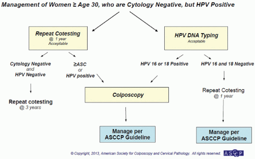
FIGURE 5.1 Management of women age 30 years and older who are cytology negative but HPV positive. (Reprinted from The Journal of Lower Genital Tract Disease, Volume 17, Number 5, with the permission of ASCCP @ American Society for Colposcopy and Cervical Pathology 2013. No copies of the algorithms may be made without the prior consent of ASCCP.)

Several assays for the detection of HPV have been developed, each with different sensitivities and specificities, depending on the method of DNA extraction from the cervical sample and the amount of DNA used. Most assays use direct hybridization techniques or nucleic acid amplification, such as the polymerase chain reaction (PCR). The two major assays currently being used to detect HPV in the United States use Hybrid Capture (HC) assay technology and include the HC II High-Risk HPV DNA test and DNA with Pap (both by Digene, Inc., Gaithersburg, MD). HC II can be performed on samples collected in the manufacturer’s media or other commercially available liquid-based cytology systems (e.g., ThinPrep by Cytyc Corp., Boxborough, MA). In 2009, Hologic produced the first test able to specifically test for HPV-16 and -18 (Cervista HPV DNA HR Test). In 2011, cobas HPV DNA 4800 (Roche) and Aptima HPV mRNA HPV (Gen-Probe) tests were Food and Drug Administration (FDA) approved.34


SCREENING FOR CERVICAL CANCER PRECURSORS

Cervical dysplasia is highly treatable, making cervical cancer almost completely preventable. For example, only 1% of treated CIN 3 will become invasive. Left untreated, however, 30% of these same lesions will become cancer over a 30-year period.35 It is estimated that 50% of women in the United States with newly diagnosed invasive cervical carcinoma have never had a Pap smear, and the other 50% occur in women who were poorly screened (e.g., had not had a Pap smear in the last 5 years), received inadequate follow-up, or were cases associated with technical errors of the Pap test process.36 Similar results have been found in Europe.37 The same could not be said in developing countries with varying technological and resource limitations. Nonetheless, attempts to improve both screening modalities and early detection of cervical cancer precursors, as well as timely intervention, remain major public health goals worldwide (Fig. 5.2).

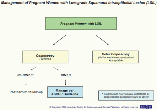
The development of new screening and management guidelines is a useful adjunct to help clinicians avoid the pitfalls of under- or overtreatment (Table 5.3 and Figs. 5.1, 5.2, 5.3, 5.4, 5.5, 5.6, 5.7, 5.8, 5.9 through 5.10). Because guidelines will likely change in the future, up-to-date information is available at http://www.uspreventiveservicestaskforce.org/uspstf/uspscerv.htm and http://www.asccp.org. Additionally, mobile device applications are available.


Liquid-Based Cytology

Although long recognized as a highly effective screening test, the validity of the conventional Pap smear has been the center of controversy due to a significant false-negative rate related in part to errors in sampling and fixation
as well as inaccurate and nonreplicable interpretation. Studies in the United Kingdom have shown a sensitivity rate of conventional Pap smears of 72% and specificity of 94%.38 Overall, about two-thirds of the false-negative Pap tests were related to sampling errors and the remaining one-third due to laboratory detection error. Liquid-based screening systems were developed to improve the transfer of cells from the collection device to the slide, provide uniformity of the cell population in each sample, decrease obscuration of menstrual blood flow, and thus decrease interpretational errors. Studies have shown liquid-based cytology to be better at providing an adequate specimen and detecting ASCUS, LSIL, and glandular abnormalities. This method is not better at detecting the clinically relevant HSIL.39, 40, 41, 42, 43


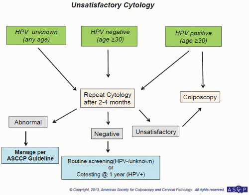
FIGURE 5.2 Unsatisfactory cytology. (Reprinted from The Journal of Lower Genital Tract Disease, Volume 17, Number 5, with the permission of ASCCP @ American Society for Colposcopy and Cervical Pathology 2013. No copies of the algorithms may be made without the prior consent of ASCCP.)

Additionally, no systematic reviews have shown liquid-based cytology to be superior overall.42,44

Added benefits of liquid-based cytology systems include the ability to perform “reflex” HPV testing and detection of other concurrent infections (gonorrhea, chlamydia, etc.). Many health care providers have therefore made liquid-based cytology their test system of choice despite higher cost. The two systems currently available are ThinPrep and SurePath (TriPath Imaging, Inc., Burlington, NC).45,46


Human Papillomavirus Testing as a Screening Modality

HPV testing (referrers to high-risk genotypes) has two benefits: increasing disease detection and increasing screening intervals. Multiple studies have shown the increased sensitivity of HPV testing compared to Pap smears especially in the cases of CIN 3 or more and adenocarcinoma in situ (AIS).47 The high sensitivity of HPV testing combined with a high negative predictive value means a negative test result should be very reassuring for patients and their health care providers. Patients younger than 21 years of age should not be screened by any modality regardless of sexual history. HPV testing as a primary screening modality in patients 21 to 29 years of age is not currently recommended because the rate of HPV infection is high in this age group. HPV testing would be more prudent as a triage modality in these patients. HPV “cotesting” (HPV with cytology) is recommended in women 30 to 65 years of age (see Table 5.3). HPV testing could also be an option in resource-poor areas because patient-collected specimens were similarly effective as Pap smears with colposcopy in a large African study.48


Human Papillomavirus Testing as a Triage Modality

For many years, a plethora of evidence has shown HPV is associated with almost 100% of high-grade dysplasia and cervical cancer. This lead to investigations, such as the ALTS (ASCUS LSIL Triage Study) trial, demonstrating that patients who are HPV negative could be managed conservatively.49 Additional research further categorized HPV into the low- and high-risk genotypes, refining our ability to triage even more. Most recently, studies have shown that HPV type 16 (HPV-16) causes 55 to 60% of cervical
cancer. HPV-18 causes 10 to 15% of cervical cancers, including a greater proportion of glandular cancers. These subtypes can be specifically tested for on the cobas HPV test or Cervista 16/18 test. The remaining 25 to 35% of cervical cancer is caused by 13 other genotypes.50, 51, 52


TABLE 5.3 Summary of Recommendations


Population


Page Number


Recommended Screening Methoda


Management of Screen Results


Comments


Age <21 years


7


No screening


HPV testing should NOT be used for screening or management of ASCUS in this age group.


Age 21-29 years


8-9


Cytology alone every 3 years


HPV-positive ASCUSb or cytology of LSIL or more severe:
Refer to ASCCP Guidelinesc
Cytology negative or HPV-negative ASCUSb:
Rescreen with cytology in 3 years


HPV testing should not be used for screening in this age group.


Age 30-65 years


9-16


HPV and cytology “cotesting” every 5 years (preferred)


HPV-positive ASCUS or cytology of LSIL or more severe:
Refer to ASCCP Guidelinesc
HPV positive, cytology negative:
Option 1—12-month follow-up with cotesting
Option 2—test for HPV-16 or HPV-16/18 genotypes
If HPV-16 or HPV-16/18 positive: refer to colposcopy
If HPV-16 or HPV-16/18 negative: 12-month follow-up with cotesting
Cotest negative or HPV-negative ASCUS:
Rescreen with cotesting in 5 years


Screening by HPV testing alone is not recommended for most clinical settings.


Cytology alone every 3 years (acceptable)


HPV-positive ASCUSb or cytology of LSIL or more severe:
Refer to ASCCP Guidelinesc
Cytology negative or HPV-negative ASCUSb:
Rescreen with cytology in 3 years


Age >65 years


16-17


No screening following adequate negative prior screening


Women with a history of CIN 2 or a more severe diagnosis should continue routine screening for at least 20 years.


After hysterectomy


17-18


No screening


Applies to women without a cervix and without a history of CIN 2 or a more severe diagnosis in the past 20 years or cervical cancer ever.


HPV vaccinated


18-19


Follow age-specific recommendations (same as unvaccinated women)


HPV, human papillomavirus; ASCUS, atypical squamous cells of undetermined significance; LSIL, low-grade intraepithelial lesions; ASCCP, American Society for Colposcopy and Cervical Pathology; CIN, cervical intraepithelial neoplasia.


a Women should not be screened annually at any age by any method.

b ASCUS cytology with secondary HPV testing for management decisions.

c From Saslow D, Solomon D, Lawson HW, et al. American Cancer Society, American Society for Colposcopy and Cervical Pathology, and American Society for Clinical Pathology screening guidelines for the prevention and early detection of cervical cancer. CA Cancer J Clin. 2012;62:147-172; Massad LS, Einstein MH, Huh WK, et al. 2012 updated consensus guidelines for the management of abnormal cervical cancer screening tests and cancer precursors. J Lower Genital Tract Dis. 2013;17(5):S1-S27.


Therefore, HPV reflex testing is especially useful for ASCUS, allowing referral to colposcopy for HPV-positive patients and surveillance for those without. The management of patients with ASCUS Pap smears has been particularly problematic because adherence with the recommended follow-up smear in 6 months tends to be poor, and many patients are lost to follow-up. This is concerning because studies have shown that patients with a diagnosis of ASCUS have a 5 to 17% chance of harboring high-grade dysplasia.53 Conversely, harm can be done (psychological, financial, physical) by further management and treatment of lesions that were destined to resolve. The ability to triage is invaluable for this cytology. HPV testing can also be used for management of women with LSIL.54 Furthermore, a patient with normal cytology and a positive HPV test can be further investigated for types 16 and 18 to determine her need for immediate colposcopy versus repeat cotesting in 1 year.55


Initiation of Screening and Screening Intervals

In 2009, the American College of Obstetrics and Gynecologists (ACOG) and the American Cancer Society
(ACS) amended its previous recommendations for screening women for cervical cancer. This was further revised in 2012 (see Table 5.3).56 The first major change involves initiation of screening. Previous guidelines recommended that women begin screening at age 21 years or 3 years after age at first coitus and that screening be continued annually until age 30 years. The current recommendations now instruct practitioners to begin screening at age 21 years and screen every 3 years thereafter until age 30 years. Initiation of screening is independent of age of first coitus and should only commence at age 21 years. In the case that an adolescent presents with known ASCUS or LSIL, cytology should be repeated in 1 year. Those that present with HSIL should
go to colposcopy. The evidence behind changing screening initiation age and interval is two-fold. First, given the prevalence of initial HPV infection in adolescents and those younger than 21 years, many young women will transiently be infected with HPV. Most of these women will clear the infection and not go on to develop cervical dysplasia. Screening before 21 years of age would therefore increase the number of surgical interventions for transient disease with virtually no risk for the development of invasive cervical cancer. Secondly, yearly screening between the ages of 21 and 30 years has not been shown to decrease the incidence of cervical cancer given the 5 to 10 years needed to develop disease.


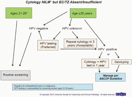
FIGURE 5.3 Cytology NILM* but EC/TZ Absent/Insufficient. (Reprinted from The Journal of Lower Genital Tract Disease, Volume 17, Number 5, with the permission of ASCCP @ American Society for Colposcopy and Cervical Pathology 2013. No copies of the algorithms may be made without the prior consent of ASCCP.)


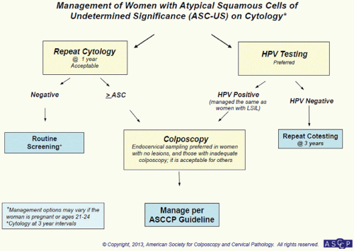
FIGURE 5.4 Management of women with atypical squamous cells of undetermined significance (ASCUS) on cytology. (Reprinted from The Journal of Lower Genital Tract Disease, Volume 17, Number 5, with the permission of ASCCP @ American Society for Colposcopy and Cervical Pathology 2013. No copies of the algorithms may be made without the prior consent of ASCCP.)


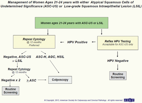
FIGURE 5.5 Management of women ages 21 to 24 years with either atypical squamous cells of undetermined significance (ASCUS) or low-grade squamous intraepithelial lesion (LSIL). (Reprinted from The Journal of Lower Genital Tract Disease, Volume 17, Number 5, with the permission of ASCCP @ American Society for Colposcopy and Cervical Pathology 2013. No copies of the algorithms may be made without the prior consent of ASCCP.)


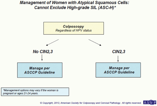
FIGURE 5.6 Management of women with atypical squamous cells: cannot exclude high-grade SIL (ASC-H). (Reprinted from The Journal of Lower Genital Tract Disease, Volume 17, Number 5, with the permission of ASCCP @ American Society for Colposcopy and Cervical Pathology 2013. No copies of the algorithms may be made without the prior consent of ASCCP.)


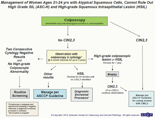
FIGURE 5.7 Management of women ages 21 to 24 years with atypical squamous cells: cannot rule out high-grade SIL (ASC-H) and high-grade squamous intraepithelial lesion (HSIL). (Reprinted from The Journal of Lower Genital Tract Disease, Volume 17, Number 5, with the permission of ASCCP @ American Society for Colposcopy and Cervical Pathology 2013. No copies of the algorithms may be made without the prior consent of ASCCP.)

The second major change to screening guidelines involves cotesting. Women younger than the age of 30 years should not be screened with HPV because the prevalence and resolution of infection is high in this group. The preferred screening for women 30 to 65 years of age is via cotesting. If negative, they can be screened every 5 years because the risk for CIN 3 or adenocarcinoma is low.


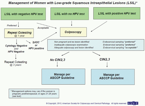
FIGURE 5.8 Management of women with low-grade squamous intraepithelial lesions (LSIL). (Reprinted from The Journal of Lower Genital Tract Disease, Volume 17, Number 5, with the permission of ASCCP @ American Society for Colposcopy and Cervical Pathology 2013. No copies of the algorithms may be made without the prior consent of ASCCP.)

A third change is management of HPV-positive, cytology-negative Pap smears in women age 30 years and older. There are two options. The practitioner can (a) repeat cotesting in 12 months or (b) order HPV genotype-specific
testing for 16/18. If HPV-16/18 are present, the patient should be immediately referred for colposcopy. If they are HPV-16/18-negative, they can be followed up in 12 months with cotesting (see Fig. 5.3).
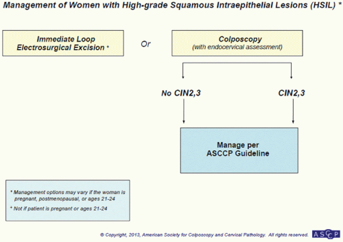


FIGURE 5.9 Management of pregnant women with low-grade squamous intraepithelial lesion (LSIL). (Reprinted from The Journal of Lower Genital Tract Disease, Volume 17, Number 5, with the permission of ASCCP @ American Society for Colposcopy and Cervical Pathology 2013. No copies of the algorithms may be made without the prior consent of ASCCP.)

The final change is the addition of the 21- to 24-year age group as a subcategory, and which includes more conservative management recommendations than those for women age 25 years and older.


FIGURE 5.10 Management of women with high-grade squamous intraepithelial lesions (HSIL). (Reprinted from The Journal of Lower Genital Tract Disease, Volume 17, Number 5, with the permission of ASCCP @ American Society for Colposcopy and Cervical Pathology 2013. No copies of the algorithms may be made without the prior consent of ASCCP.)

Only gold members can continue reading. Log In or Register to continue

Stay updated, free articles. Join our Telegram channel

Jun 25, 2016 | Posted by in GYNECOLOGY | Comments Off on Abnormal Cervical Cytology and Human Papillomavirus

Full access? Get Clinical Tree

Get Clinical Tree app for offline access