Abdominal pain and vomiting

20.1 Abdominal pain and vomiting


Acute abdominal pain and vomiting are common symptoms in children and a frequent reason for children to be taken to the doctor. Their causes are many and diverse; those that require surgery must be distinguished from those with a medical origin. Although there is considerable overlap of age in many disorders, other conditions occur only within a specific age range; for example, pyloric stenosis is not seen after the age of 3 months.


Abdominal pain later in the first year


The main surgical cause of abdominal pain between 3 and 12 months of age is intussusception. Vomiting is a frequent accompanying feature, such that, when the colicky abdominal pain is not pronounced, intussusception must be distinguished from other causes of vomiting in this age group (see below).


Intussusception


In intussusception, the distal ileum (the intussusceptum) telescopes into adjoining distal bowel (the intussuscipiens), resulting in intestinal obstruction. It can occur at any age but is most likely in the infant between 3 and 18  months who suddenly develops screaming attacks of pain with vomiting. During each episode of pain the infant becomes pale and may draw up the legs.


The spasms of pain tend to last for 2–3 minutes and occur at intervals of about 10–20 minutes, although after a while the pain becomes more persistent. Vomiting is an early symptom. The passage of a few loose stools early on represents evacuation of the bowel distal to the obstruction. The small volume and limited duration of loose stools in intussusception helps differentiate it from acute gastroenteritis. Congestion of the intussusceptum may lead to the passage of blood-stained or ‘redcurrant jelly’ stools. Many infants with intussusception present with little more than pallor, lethargy and vomiting, and may have little evidence of abdominal pain. Should these symptoms be ignored, the infant may progress to develop signs of septicaemia or shock.


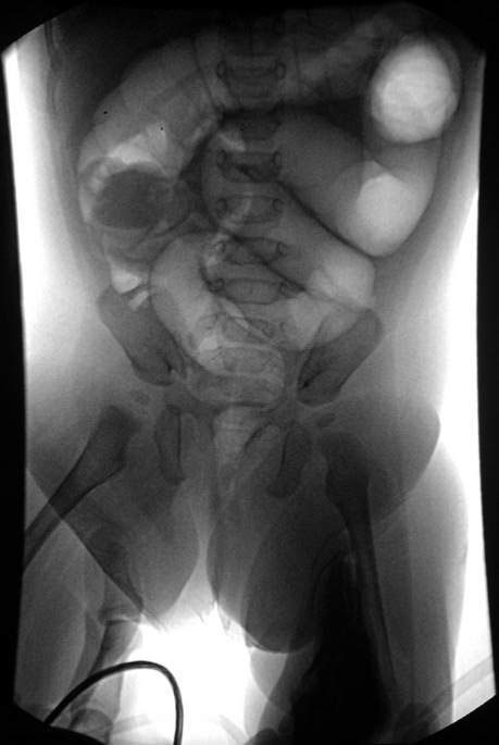
The infant with intussusception looks pale, lethargic, anxious and unwell. A vague mass may be felt in the right or left upper quadrants of the abdomen but, once abdominal distension has developed, the mass becomes obscure and difficult to palpate (Fig. 20.1.1). The apex of the intussusceptum may be palpable on rectal examination in a few, and the examining glove may be blood-stained. A plain X-ray of the abdomen will often be normal but may show an unusual bowel gas distribution or features of bowel obstruction. Ultrasound examination may be helpful in making the diagnosis. Where intussusception is suspected clinically or confirmed on ultrasonography, a gas or barium enema must be performed (unless the child has peritonitis). The enema will demonstrate the position of the apex of the intussusception (Fig. 20.1.2).


Acute abdominal pain in older children


Children often present with abdominal pain and in most no specific cause is found. Constipation and mesenteric adenitis are probably the most common non-surgical identifiable causes.


Acute appendicitis


Appendicitis may occur at any age, although it is rare under 5  years of age. Early diagnosis is difficult in the young child (under 5  years) and in developmentally delayed children; many of these children have established peritonitis or an appendix abscess at presentation. Delays in the diagnosis of acute appendicitis in childhood is related in part to its variable symptomatology. For example, there may be relatively little abdominal pain, vomiting may be absent and diarrhoea may be a misleading feature.


Nevertheless, the most important and consistent feature is localized abdominal pain. The pain may be intermittent and colicky initially, or situated in the epigastrium or periumbilical region, but soon shifts to the right iliac fossa. Constant pain that is worse with movement is the result of peritoneal irritation (‘peritonism’). Vomiting occurs in the majority of children, and some may pass a loose stool. The temperature is usually normal or slightly raised, but occasionally may be in excess of 38 °C.


Physical examination of the abdomen should be directed at showing that movement of adjacent peritoneal surfaces exacerbates the pain. The child’s cooperation makes assessment easier, and repeated examination of the abdomen may be required to make the diagnosis. A child with appendicitis usually will exhibit tenderness and guarding localized to the right iliac fossa. Gentle palpation and percussion tenderness, performed while observing the child’s face, will provide the most reliable evidence of abdominal tenderness and involuntary guarding. Rebound tenderness is an unreliable sign in children, and attempts to elicit the sign may cause unnecessary pain and destroy the child’s confidence in the doctor. Rectal examination is required rarely and is primarily indicated if a pelvic appendix or pelvic collection is suspected. It should not be performed when examination of the ventral abdominal wall has already enabled a confident diagnosis of acute appendicitis to be made. Bowel sounds may be normal or reduced and contribute little to the diagnosis.


Peritonitis should be suspected when the child is acutely ill with abdominal pain and fever and is reluctant to move. On examination, there will be generalized abdominal tenderness and guarding.


Laboratory studies are rarely helpful in making the diagnosis but the urine should be checked routinely. Ultrasonography has a role when the diagnosis is uncertain.


Differential diagnosis


Mesenteric adenitis is the most difficult disorder to distinguish from acute appendicitis. In general, localization of pain and tenderness is variable and less specific, and the temperature may be higher. Guarding is rarely present in mesenteric lymphadenitis.


Other conditions that may mimic acute appendicitis are relatively uncommon. Meckel diverticulitis has symptoms identical to those of appendicitis, such that differentiation is possible only at laparoscopy or laparotomy. Pain in the right iliac fossa may represent radiation from torsion of the right testis or a strangulated inguinal hernia, and highlights the importance of examination of the genitalia in all boys with lower abdominal symptoms (see Chapter 9.1). Acute abdominal pain may occur with renal colic, pyelonephritis and, at times, acute glomerulonephritis. Pain and tenderness is usually referred to the loin. Urine analysis and radiology will confirm the diagnosis. In Henoch–Schönlein purpura, the abdominal pain is often severe and colicky, and may be accompanied by vomiting. The characteristic skin lesions over the buttock and legs may be inconspicuous or absent when the child is first examined.


In the appropriate ethnic group, sickle cell anaemia is a prominent cause of acute abdominal pain and should be considered in a pale child with splenomegaly.


Children with cystic fibrosis frequently experience episodes of abdominal pain from faecal impaction (called ‘meconium ileus equivalent’), a well-known manifestation of this disease. The symptoms resolve following a bowel washout.


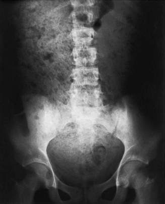
It is unusual for constipation in an otherwise normal child to produce sufficient abdominal pain to suggest a surgical emergency. A plain X-ray of the abdomen will demonstrate the extent of faecal accumulation (Fig. 20.1.3). It should be remembered, however, that the diagnosis of constipation can almost exclusively be made on clinical grounds. Abdominal X-ray is not a standard tool in assessing for constipation and should be reserved solely for other indications or more complex cases.


Stay updated, free articles. Join our Telegram channel

Aug 4, 2016 | Posted by in PEDIATRICS | Comments Off on Abdominal pain and vomiting

Full access? Get Clinical Tree

Get Clinical Tree app for offline access