Abdominal Mass in Neonate
Eva Ilse Rubio, MD
DIFFERENTIAL DIAGNOSIS
Common
Urinary Tract Obstruction
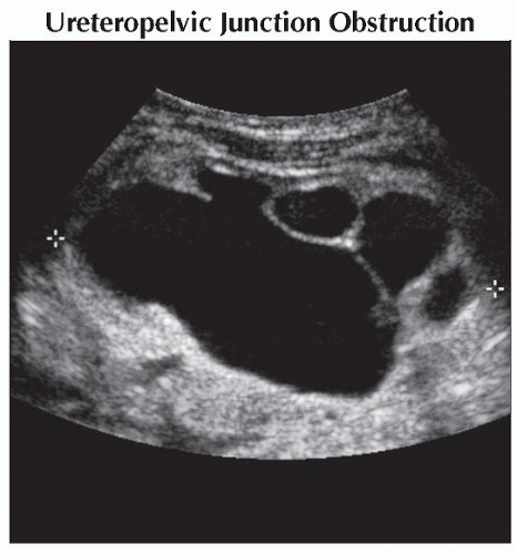
Ureteropelvic Junction Obstruction
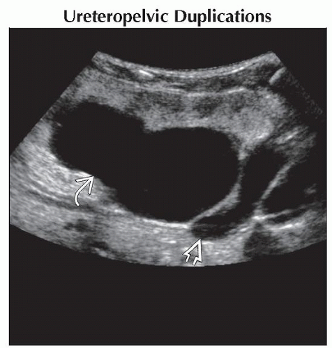
Ureteropelvic Duplications
Multicystic Dysplastic Kidney
Neuroblastoma, Congenital
Adrenal Hemorrhage
Gastrointestinal Duplication Cyst
Mesenteric Lymphatic Malformations
Mesoblastic Nephroma
Teratoma
Less Common
Ovarian Torsion
Hepatoblastoma
Meconium Pseudocyst
Hemangioendothelioma
Vascular Malformation
Pulmonary Sequestration
Rare but Important
Hydrocolpos/Hydrometrocolpos
Wilms Tumor (Congenital)
ESSENTIAL INFORMATION
Key Differential Diagnosis Issues
Male vs. female
Other clinical findings: Skin lesions, urinary output, meconium passage
Cystic, solid, or mixed cystic/solid
Presence or absence of calcifications
Helpful Clues for Common Diagnoses
Urinary Tract Obstruction
Ureteropelvic junction obstruction: Larger cystic, anechoic renal pelvis surrounded by smaller cystic, anechoic calyces
Most common congenital obstruction of urinary tract
Hydronephrosis without hydroureter
Sometimes difficult to differentiate from multicystic dysplastic kidney (MCDK)
Abnormalities common in contralateral kidney: Reflux or ureteropelvic junction obstruction
Duplication with obstructed upper pole appear as complex or simple cystic structure
Reflux common in lower pole ureter
Diagnosis is often bilateral but asymmetric in severity
Multicystic Dysplastic Kidney
Most common appearance is noncommunicating cysts of varying sizes; usually spontaneously involute over time
Variable appearance depending on stage
May be massive and bizarre
Large enough to cause respiratory distress in rare cases
May have residual intervening dysplastic renal parenchyma
± identifiable ureter
Timing to complete involution varies from prenatal period to late teens
Higher incidence of other genitourinary abnormalities
Neuroblastoma, Congenital
Origin in sympathetic chain ganglia
Occurs anywhere from coccyx to skull base
Vast majority arise in adrenal gland
Often heterogeneous, mixed cystic and solid
Coarse calcifications are common
Assess for vertebral involvement, spinal canal invasion, rib splaying
Engulfs large vessels
Mass often elevates aorta from spine
Adrenal Hemorrhage
May be large and difficult to differentiate from neuroblastoma
No internal blood flow detected on US
Involutes over time
Usually circumscribed, heterogeneous, often with cystic components
Gastrointestinal Duplication Cyst
Usually round structure
± bowel “wall signature” of hypoechoic wall/mucosa layers
Hypoechoic or echogenic contents
Usually does not communicate with GI lumen
Most common locations are terminal ileum and esophagus
Other GI locations are uncommon
Mesenteric Lymphatic Malformations
Nonspecific hypoechoic/anechoic structure
May have irregular or lobulated borders
May have septations
Mesoblastic Nephroma
Most common neonatal renal neoplasm
Encapsulated calcifications rare
Solid, may be heterogeneous
US: Whorled, heterogeneous, compared to uterine fibroid
CT: Solid, with mild enhancement
MR: Intermediate on T1, bright on T2
Usually benign; however, beware small subset that may recur or metastasize
Helpful Clues for Less Common Diagnoses
Ovarian Torsion
May be suspected if only 1 ovary can be identified
Circumscribed, heterogeneous pelvic mass; may see peripheral follicles
May be precipitated by dominant cyst or underlying mass, such as teratoma
Pitfall: Demonstration of blood flow by US may confound diagnosis due to intermittent torsion or multiplicity of blood supply to ovaries
Meconium Pseudocyst
Radiographically, large soft tissue density with mass effect
Lesional or peritoneal calcifications may have wispy or “eggshell” appearance
Distal bowel obstruction pattern typical
Hemangioendothelioma
Variable appearance; most commonly multiple focal, round, target-like lesions within liver
May present as large solitary lesion
Highly vascular lesion
Great vessels distal to lesion (aorta, superior mesenteric artery) may be attenuated
Look for signs of congestive heart failure
Vascular Malformation
Highly variable appearance, depending on histology, presence of necrosis, stage of involution
Helpful Clues for Rare Diagnoses
Hydrocolpos/Hydrometrocolpos
Often associated with other anomalies
Cloaca, urogenital sinus, renal agenesis, cystic kidneys, esophageal/duodenal atresia, anorectal malformation
Occasionally seen in intersex conditions
Wilms Tumor (Congenital)
Cannot be differentiated from mesoblastic nephroma by imaging
Associated with other conditions
Beckwith-Wiedemann: Hypertrophy
WAGR: Wilms, aniridia, genitourinary anomalies, mental retardation
Drash: Pseudohermaphroditism, renal failure
Tends to invade vessel lumen (renal vein, IVC) rather than surround/engulf vessels (as seen with neuroblastoma)
Contralateral kidney risks: Bilateral Wilms, nephroblastomatosis
Wilms tumor nearly always occurs after 1st year of life; congenital/neonatal presentation extremely uncommon
Image Gallery
 Longitudinal ultrasound shows a markedly dilated upper pole
 with portions of the dilated, tortuous upper pole ureter with portions of the dilated, tortuous upper pole ureter  also visualized. A ureterocele was the cause of the upper pole obstruction. also visualized. A ureterocele was the cause of the upper pole obstruction.Stay updated, free articles. Join our Telegram channel
Full access? Get Clinical Tree
 Get Clinical Tree app for offline access

|
