Abdominal Mass in a Child
Alexander J. Towbin, MD
DIFFERENTIAL DIAGNOSIS
Common
Hydronephrosis
Splenomegaly
Appendiceal Abscess
Wilms Tumor
Neuroblastoma
Less Common
Rhabdomyosarcoma
Ovarian Tumors
Multicystic Dysplastic Kidney
Hemangioendothelioma/Hemangioma
Hepatoblastoma
Mesoblastic Nephroma
Hepatocellular Carcinoma
Rare but Important
Renal Cell Carcinoma
Pancreatoblastoma
ESSENTIAL INFORMATION
Key Differential Diagnosis Issues
> 50% of abdominal masses are renal
Hydronephrosis and multicystic dysplastic kidney are most common in neonates
Hydronephrosis and Wilms tumor are most common in infants and children
Neuroblastoma and Wilms tumor account for majority of abdominal malignancies
Helpful Clues for Common Diagnoses
Hydronephrosis
Most common abdominal mass
Diagnosed in 1-5% of pregnancies
Up to 30% are bilateral
Resolves on postnatal US in ˜ 50%
10% have UPJ obstruction
Vesicoureteral reflux in 10%
Postnatal US should be 1st imaging test
Hint: In males with bilateral hydronephrosis, consider posterior urethral valves
Splenomegaly
Many causes of splenomegaly in children
Common causes include infection, right heart failure, and leukemia/lymphoma
May appear as mass on radiograph with displacement of bowel and stomach
Appendiceal Abscess
Seen after ruptured appendix
Occurs in ˜ 4% of cases
More common in children < 4 years
Often have symptoms more than 3 days
Wilms Tumor
Most common abdominal neoplasm
2nd most common pediatric solid tumor
Occurs in children < 15 years of age with peak at 3 years
May be bilateral
Associated with WAGR, Denys-Drash syndrome, and Beckwith-Wiedemann
2x more common with horseshoe kidney
Appears as heterogeneous renal mass
Calcifications in 15-20%
Typically displaces vessels ± inferior vena cava invasion
Neuroblastoma
Most common pediatric solid tumor
6-10% of all childhood cancers
˜ 15% of cancer-related deaths in children
˜ 30% occur in 1st year of life with peak at 0-4 years
2nd most common abdominal tumor
˜ 65% arise in abdomen
Usually arise from adrenal medulla
Can arise anywhere along sympathetic chain
Usually presents as asymptomatic abdominal mass
Metastases present in 70% at diagnosis
Bone and bone marrow
Liver metastases can be focal or diffuse
Urine catecholamines are elevated in 90%
I-123 MIBG positive in 90%
Calcifications present in ˜ 85% on CT
Typically encases vessels
Helpful Clues for Less Common Diagnoses
Rhabdomyosarcoma
Most common pediatric soft tissue sarcoma
Accounts for 5% of pediatric cancers
GU origin common for abdominal tumors
Bladder is most common organ of origin
Usually presents before age 5
Metastases to lung and marrow
Ovarian Tumors
Teratomas are most common tumor
Majority are benign
Malignant ovarian tumors account for 1% of pediatric cancers
75-80% are germ cell tumors
Can become very large and extend to abdomen
Multicystic Dysplastic Kidney
More common in males
Left kidney more commonly affected
Natural history is involution of kidney
Distinguished from hydronephrosis as cysts do not connect with renal pelvis
Hemangioendothelioma/Hemangioma
a.k.a. infantile hepatic hemangioma
Most common benign hepatic tumor
85% diagnosed in 1st 6 months
Skin hemangiomas present in ˜ 50%
Can present with high-output heart failure
Hepatoblastoma
Most common pediatric liver malignancy
1% of all pediatric malignancies
79% of liver malignancies < 15 years
Most diagnosed under 18 months of age
Associated with low birth weight, hemihypertrophy, Beckwith-Wiedemann, familial adenomatous polyposis, trisomy 18, and fetal alcohol syndrome
90% have increased serum AFP
Most common in right lobe of liver
Calcifications in 40-55%
Distant metastases in 20% at diagnosis
Lung, brain, and bone
Mesoblastic Nephroma
3-10% of pediatric renal tumors
Most common renal tumor in infants, 90% diagnosed in 1st year
2x more common in males
Predominantly solid with variable cystic areas
Hepatocellular Carcinoma
2nd most common pediatric liver malignancy
Rare before age 5
˜ 75% are not associated with liver disease
Risks: Preexisting cirrhosis due to biliary atresia, Fanconi syndrome, viral hepatitis, hereditary tyrosinemia, or glycogen storage disease
Other risks: Androgen steroids, oral contraceptives, methotrexate
Metastases common at diagnosis
Regional lymph nodes, lungs, bone
Elevated AFP in 60-80%
Helpful Clues for Rare Diagnoses
Renal Cell Carcinoma
Accounts for 2-5% of pediatric renal tumors
Mean age 9-15 years
Metastases in 20% at diagnosis
Associated with von Hippel-Lindau disease
Pancreatoblastoma
Most common pediatric pancreatic neoplasm
Neoplasm of acinar cells
Most common in 1st decade of life
Associated with Beckwith-Wiedemann
AFP elevated in up to 55%
Usually large solitary pancreatic mass
Well defined with lobulated margins
˜ 50% in pancreatic head
Image Gallery
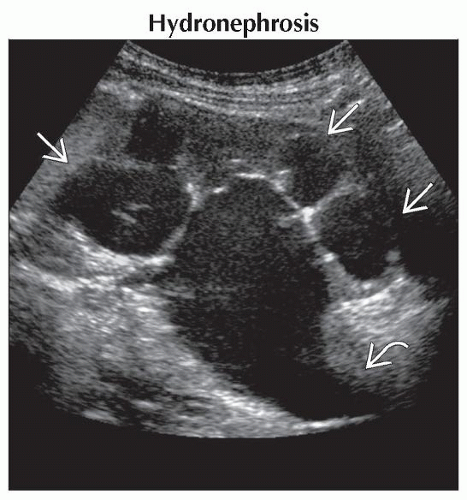 Longitudinal ultrasound shows marked hydronephrosis  and a dilated proximal ureter and a dilated proximal ureter  . The more distal ureter was not seen. Obstruction of the ureteropelvic junction is a common cause of hydronephrosis. . The more distal ureter was not seen. Obstruction of the ureteropelvic junction is a common cause of hydronephrosis. |
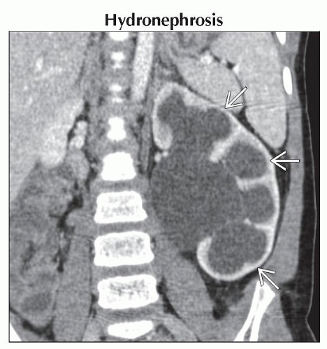 Coronal CECT shows marked hydronephrosis of the left kidney and associated cortical thinning
 . The distal ureter was not seen. The patient was asymptomatic despite this congenital abnormality. . The distal ureter was not seen. The patient was asymptomatic despite this congenital abnormality.Stay updated, free articles. Join our Telegram channel
Full access? Get Clinical Tree
 Get Clinical Tree app for offline access

|