Abdominal Calcifications
Michael Nasser, MD
DIFFERENTIAL DIAGNOSIS
Common
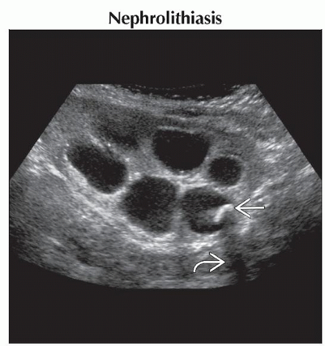
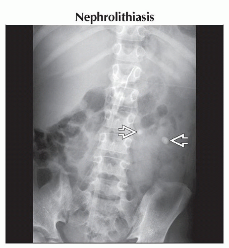
Nephrolithiasis
Cholelithiasis
Hepatic and Splenic Granulomas
Neuroblastoma
Hepatoblastoma
Teratoma (Ovarian)
Appendicolith
Less Common
Remote Adrenal Hemorrhage
Meconium Peritonitis
ESSENTIAL INFORMATION
Key Differential Diagnosis Issues
Age of patient
Location of calcification
Morphology of calcifications: Coarse, punctate, or curvilinear
Helpful Clues for Common Diagnoses
Nephrolithiasis
Conventional radiographs show punctate or coarse calcification that projects over
Renal shadow
Course of ureters
Expected location of bladder
Renal calculi are echogenic with posterior shadowing on ultrasound
Color Doppler may be used to look for “twinkling” artifact or “comet tail” artifact posterior to small renal stones
Cholelithiasis
˜ 30% of gallstones are detectable by plain film
Look for calcification projecting over medial aspect of hepatic silhouette on conventional radiographs
US is preferred modality for evaluation
Gallstones are echogenic with posterior shadowing
Hepatic and Splenic Granulomas
Multiple, punctate, round, or ovoid-shaped calcifications projecting over spleen &/or liver on plain film
Appear as multiple, punctate, echogenic foci on ultrasound that may or may not have posterior shadowing
Neuroblastoma
Can arise anywhere along sympathetic chain from skull base to pelvis
Median age at diagnosis is 2 years old
Involvement of adrenal glands and extraadrenal retroperitoneum make up more than 60% of cases
Conventional radiographic findings
Paraspinal soft tissue mass
30% will have associated calcifications
May have lytic, sclerotic, or mixed metastatic bone lesions
CT findings
Soft tissue mass with heterogeneous attenuation (necrosis &/or hemorrhage)
Associated calcifications in up to 85% of cases
Engulfs adjacent vascular structures, such as celiac and superior mesenteric artery
Metastasis most common to liver & bone
Bone metastasis may be lytic, sclerotic, or mixed pattern
Look for involvement of neuroforamina
MR findings: Best for detecting intraspinal extension of tumor
Hepatoblastoma
Majority occur in children < 3 years old
Painless abdominal mass
Elevated α-fetoprotein levels
Conventional radiographic findings
Large, soft tissue mass in right upper quadrant of abdomen
May displace bowel
Roughly 1/2 will have visible calcifications, usually coarse
CT findings
Usually large at presentation
Well-defined lesion
> 60% located in right lobe of liver
Heterogeneous in attenuation due to necrosis and hemorrhage
Heterogeneous enhancement
Roughly 50% have coarse calcifications
CT of chest is performed for metastatic workup
Teratoma (Ovarian)
Conventional radiographic findings
Punctate or coarse calcifications projecting over pelvis
May see associated “teeth” within pelvis
Mass effect upon adjacent structures
Ultrasound findings
Heterogeneous echogenicity
Solid and cystic components
Fat and hair are echogenic
Calcifications are echogenic with posterior shadowing
May contain fat-fluid levels
Ovarian torsion may be a complication
CT findings
Solid and cystic components
May have fluid levels
May contain fat
May contain coarse calcifications
May be bilateral in up to 15% of cases
Appendicolith
10-15% of acute appendicitis associated with appendicolith
Appendicolith may be round or oval-shaped
Usually in right lower abdomen/pelvis
May be in right upper abdomen in retrocecal appendix
Conventional radiographic findings
Appendicolith (10-15%)
Air-fluid levels localized to right lower abdomen
Splinting (levocurvature of lower spine)
CT findings
Appendicolith
Appendiceal diameter > 6 mm
Fluid-filled appendix with enhancement of appendiceal wall
Periappendiceal soft tissue infiltration
Look for abscess and extraluminal appendicolith with perforation
Helpful Clues for Less Common Diagnoses
Remote Adrenal Hemorrhage
Resolving adrenal hemorrhage may calcify
Secondary to any stress in perinatal period
Bilateral in 10% of cases
Punctate calcification may be seen on conventional radiographs projecting over suprarenal region
Noncontrast abdominal CT to confirm intra-adrenal location
Ultrasound with Doppler helpful to show lack of flow vs. flow in neuroblastoma
Meconium Peritonitis
Sterile chemical reaction resulting from bowel perforation in utero
1 in 35,000 live births
Conventional radiographic findings
Diffuse calcification
Sometimes pseudocyst formation (peripheral calcifications)
May displace bowel
Ultrasound findings
May be diagnosed in utero during 2nd or 3rd trimester
Diffuse hyperechoic punctate echoes with or without acoustic shadowing
Especially along hepatic surface & in scrotum
May have ascites, polyhydramnios &/or bowel distention
If pseudocyst formation: Cystic heterogeneous mass with an irregular, calcified wall
Image Gallery
 Longitudinal ultrasound shows an echogenic
 , shadowing , shadowing  renal stone. The associated hydronephrosis of the kidney suggests that there is an obstructing renal stone distally in the collecting system. renal stone. The associated hydronephrosis of the kidney suggests that there is an obstructing renal stone distally in the collecting system.Stay updated, free articles. Join our Telegram channel
Full access? Get Clinical Tree
 Get Clinical Tree app for offline access

|

