Vulvovaginitis, Sexually Transmitted Infections, and Pelvic Inflammatory Disease

Chapter 22 Vulvovaginitis, Sexually Transmitted Infections, and Pelvic Inflammatory Disease


Infectious diseases are interesting and challenging disorders in gynecology. Some are difficult to treat and have frequent recurrences. Antibiotic resistance has recently changed the recommended treatment for one of the original “venereal diseases,” and vaccination has been introduced as a preventive tool for one of the more prevalent pelvic infections. The diagnosis and management of common reproductive tract infections in nonpregnant women are covered in this chapter.


image Normal Physiology and Microecology of the Vagina


The vagina is lined by nonkeratinized stratified squamous epithelium, which is powerfully influenced by estrogen and progesterone. The vagina of the newborn is colonized by aerobic and anaerobic bacteria acquired while passing through the birth canal. The newborn’s vaginal epithelium is strongly estrogenized and rich in glycogen, which supports growth of lactic acid–producing lactobacilli. This results in a low pH (<4.7), which promotes further growth of acidophilic-protective microflora. Within days of birth, estrogen decreases, and the vaginal epithelium becomes thin, atrophic, and largely devoid of glycogen. In this environment, the pH rises, and acidophilic organisms no longer have a selective advantage. As a consequence, the predominant vaginal microflora becomes diverse gram-positive cocci and bacilli.


With the onset of puberty and ovarian steroidogenesis, the vagina again becomes estrogenized, and the glycogen content increases. Lactic acid– and hydrogen peroxide (H2O2)–producing lactobacilli again predominate, resulting in a self-sustaining vaginal pH of between 3.5 and 4.5. Even so, a wide variety of aerobic and anaerobic bacteria can be cultured from the normal vagina. Most women harbor at least three to eight types of bacteria at any given time. Lactic acid, H2O2, and other substances produced by “healthy” lactobacilli provide some protection in the lower reproductive tract from STIs, including human immunodeficiency virus (HIV).


Multiple factors may alter this protective microflora. Antibiotics suppress growth of commensal organisms, which can allow pathogenic strains (e.g., yeasts) to predominate. Douching with water or nonbuffered solutions may transiently alter the pH or selectively suppress endogenous bacteria. Sexual intercourse with introduction of semen raises the pH to as high as 7.2 for 6 to 8 hours, making the vaginal milieu receptive to STI pathogens. During coitus, vaginal transudate increases vaginal fluid and serves as lubrication. Vaginal transudate has the same pH as blood (7.4), which also favors attachment of abnormal microflora. The presence of a foreign body (e.g., forgotten diaphragm or tampon in adults or various small objects in children) dramatically disrupts normal vaginal cleansing mechanisms and may lead to secondary infection.


INVESTIGATION OF VAGINAL DISCHARGE


Patients with a vaginal infection frequently complain of a nonbloody vaginal discharge. The characteristics of the discharge (e.g., color, texture, viscosity, and odor) are helpful in making the correct diagnosis. Normal vaginal fluid has a pH of less than 4.7 in ovulatory women. Vaginal pH can be easily determined by using pH paper with an appropriate pH range (3.5 to 7.0). Self-care over-the-counter (OTC) tests, such as “pH on a stick” and “pH glove,” are available to test vaginal pH.


Testing for the presence of an amine odor (referred to as a positive whiff test) is performed by putting a few drops of 10% potassium hydroxide (KOH) in the spoon of a vaginal speculum after it is removed from the vagina. With healthy vaginal fluid, no odor is noted. An “amine” or “fishy” odor suggests trichomoniasis or bacterial vaginosis (BV).


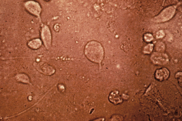
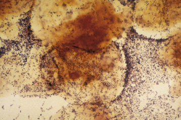
A wet-mount preparation of the discharge should be evaluated. Using a cotton-tipped applicator, a sample of vaginal discharge from the posterior fornix is suspended in 2 mL of normal saline. A drop of this solution is placed on a glass slide, covered with a coverslip, and examined under the microscope. Motile trichomonads may be seen on this type of wet mount (Figure 22-1). Also, epithelial cells with irregular, granular edges (clue cells) are indicative of clumped bacteria on the cell wall and are highly suggestive of BV if present in more than 20% of epithelial cells (Figure 22-2). If the cells are not sufficiently separated, an aliquot of fluid is placed in a drop of saline or 10% to 20% KOH (to eliminate cellular and other debris while leaving mycelia) and examined microscopically, to visualize the pseudohyphae or spores of Candida infection. In complicated or atypical cases, bacterial and yeast cultures are required.


image Vaginal Discharge Etiology


Vaginitis and complaints referable to the vagina (e.g., discharge, pruritus, burning, and “late” dysuria) are common. A correct diagnosis can be difficult because symptoms and signs may be nonspecific, patients frequently self-medicate with OTC products, multiple causes may be present, and accurate diagnostic tools are either not available or not properly employed.


Up to 90% of cases of vaginitis appear to be caused by three conditions. Bacterial vaginosis accounts for 40% to 50%, vulvovaginal candidiasis (VVC) for 20% to 25%, and trichomoniasis for 15% or less of cases. Mucopurulent cervicitis (“mucopus”) caused by chlamydia, Neisseria gonorrhoeae, mycoplasma, or BV-associated bacteria (see Figure 22-6) may also cause vaginal irritation and discharge. Less common types include atrophic vaginitis (overgrowth with aerobic or anaerobic microflora in the absence of lactobacilli and with hypoestrogenized tissues), foreign-body vaginitis, genital ulcer diseases such as herpes and syphilis, desquamative vaginitis (most commonly group B streptococcal overgrowth), and lichen planus. Irritation from sexual activity and allergen-containing substances can also mimic infectious vaginitis.


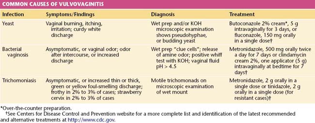
Standard clinic or office diagnosis of vaginitis requires a working microscope, pH paper, KOH, saline solution, slides, coverslips, and the ability to recognize an amine odor (whiff test). In many settings, these rudimentary tools are not available. Newer, inexpensive “point of care” products can detect vaginal sialidase, amines, and proline aminopeptidase and other biomarker substances or nucleic acid–based tests. In difficult or refractory cases, additional tests for vaginal agents may be used such as culture for Trichomonas vaginalis, Candida, mycoplasmas, or predominant vaginal aerobic bacterial. Table 22-1 lists the common causes, characteristics, and treatments for vulvar and vaginal infections.


VULVOVAGINAL CANDIDIASIS


VVC is the second most common cause of vulvovaginal-related symptoms. Wide use of OTC antifungal products for self-diagnosed candidiasis has resulted in a decreased frequency of doctor visits, but women who do present are less likely to present with textbook findings. Candida albicans formerly caused more than 90% of cases of VVC, but now less azole-susceptible species, such as Candida glabrata, are recognized as causative agents in 15% of cases. These less susceptible yeasts necessitate prolonged or alternative treatments. Because of Candida’s requirement for estrogenated tissues, VVC becomes more common after menarche and less common after menopause. An estimated 75% of women acquire VVC at some time in their life, and 5% suffer frequent symptomatic recurrences. VVC is considered recurrent when at least four episodes occur within 1 year. Risk factors for recurrent VVC include high-dose oral contraceptives, diaphragm use with a spermicide, diabetes mellitus, antibiotic use, pregnancy, immunosuppression from any cause (HIV/AIDS, transplantation, steroid use), and possibly tight occlusive clothing.


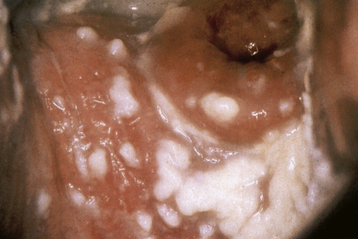
The classic presentation of VVC includes vaginal itching, burning, irritation, and possibly postvoiding dysuria. The discharge is usually odorless, has a pH of less than 4.7, and is thick or curdy with the appearance of cottage cheese (Figure 22-3). Examination often shows vulvovaginal erythema, with evidence of acute or chronic excoriation.


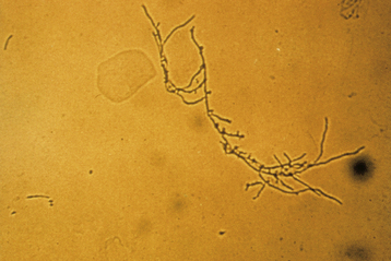
Microscopic examination of a wet-mount preparation is positive for budding yeast cells, pseudohyphae, or mycelial tangles (Figure 22-4) in 50% to 70% of cases. Women with suggestive clinical findings but absent wet preparation evidence may benefit from fungal culture.


First-line treatments include topical or oral antifungal (imidazole) agents. Documented cases of recurrences can be effectively treated by confirming the diagnosis and treating with weekly suppressive doses of topical imidazoles. Boric acid (600 mg vaginal gelatin capsules) 3 times daily for 1 week is an effective treatment for imidazole-resistant species. Although VVC is not thought to be sexually transmitted in most cases, male partners sometimes reinfect their partners and are routinely treated by some practitioners.


TRICHOMONIASIS


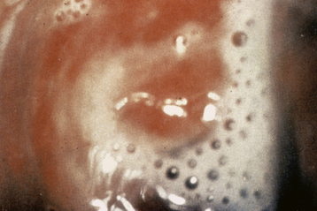
Trichomoniasis (cervicitis, vaginitis, and urethritis) is caused by the protozoan T. vaginalis. About 50% of cases in women and men are asymptomatic. Symptomatic infection is classically manifested by a green-yellow, frothy vaginal discharge (Figure 22-5), with a “musty” odor. Dyspareunia, vulvovaginal irritation, and occasionally dysuria may be present. Male partners are often asymptomatic even though they demonstrate nongonococcal urethritis on direct examination.


The diagnosis of trichomoniasis in patients and their sexual partners should be followed by screening for other prevalent STIs and empiric treatment of partners. Diagnosis is usually made on clinical grounds and can be confirmed by seeing the characteristic motility of trichomonads on a saline wet mount. Much more sensitive techniques, including culture, polymerase chain reaction (PCR), and antigen testing, are becoming available.


In addition to vulvovaginitis and urethritis, trichomoniasis is associated with upper reproductive tract symptoms, an increased risk for adverse pregnancy outcomes (prematurity, low birth weight), and increased transmission of HIV infection.


Metronidazole (2 g single oral dose) is the recommended treatment (see Table 22-1). Patients should not consume alcohol for 2 days after treatment. Although metronidazole is not known to be teratogenic in recommended dosages, it has been traditionally avoided during the first 12 weeks of pregnancy. Prompt early treatment during pregnancy relieves symptoms, reduces the risk for HIV transmission, and may improve pregnancy outcomes. Trichomoniasis should be treated before vaginal surgical procedures.


Metronidazole resistance is increasing and may be overcome by using tinidazole (now available in the United States; see Table 22-1) or using higher doses of metronidazole (2 g daily for 7 days). Reversible side effects of metronidazole include an “Antabuse-like reaction” with alcohol exposure, neutropenia, and peripheral neuropathy. Higher-dose treatment for resistant trichomoniasis in pregnancy should be prescribed with caution in consultation with experts.


Jun 4, 2016 | Posted by in GYNECOLOGY | Comments Off on Vulvovaginitis, Sexually Transmitted Infections, and Pelvic Inflammatory Disease

Full access? Get Clinical Tree

Get Clinical Tree app for offline access