Chapter 40 Vulvar and Vaginal Cancer
Vulvar Neoplasms
Malignant tumors of the vulva are uncommon, representing about 4% of malignancies of the female genital tract. Most tumors are squamous cell carcinomas, with melanomas, adenocarcinomas, basal cell carcinomas, and sarcomas occurring much less frequently.
Squamous cell carcinoma of the vulva occurs mainly in postmenopausal women, and the mean age at diagnosis is 65 years. A history of chronic vulvar itching is common.
EPIDEMIOLOGY
Recent studies suggest two different etiologic types of vulvar cancer. One type is seen mainly in younger patients, is related to human papillomavirus (HPV) infection and smoking, and is commonly associated with vulvar intraepithelial neoplasia (VIN) of the basaloid or warty type. The more common type is seen mainly in elderly women and is unrelated to smoking or HPV infection; concurrent VIN is uncommon, but long-standing lichen sclerosus is common. When VIN is present, it is of the differentiated type. VIN III carries a significant risk for progression to invasive cancer if left untreated.
About 5% of patients have positive results on serologic testing for syphilis. In the latter group of patients, vulvar cancer occurs at an earlier age and carries a graver prognosis. Although rarely seen in the United States, vulvar cancer also occurs in association with lymphogranuloma venereum and granuloma inguinale.
Intraepithelial Neoplasia
The International Society for the Study of Vulvar Disease recognizes two varieties of intraepithelial neoplasia: squamous cell carcinoma in situ (Bowen’s disease) or VIN III, and Paget’s disease. With the introduction of the HPV vaccines, there should be a significant reduction in the incidence of VIN and invasive vulvar cancer, particularly in young patients, in the future.
SQUAMOUS CELL CARCINOMA IN SITU: VULVAR INTRAEPITHELIAL NEOPLASIA TYPE III
During the past 25 years, the incidence of VIN has increased markedly. Younger patients are being affected, and the mean age is about 45 years.
Clinical Features
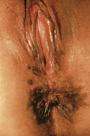
Itching is the most common symptom, although some patients present with palpable or visible abnormalities of the vulva. About half of patients are asymptomatic. There is no absolutely diagnostic appearance. Most lesions are elevated, but the color may be white, red, pink, gray, or brown (Figure 40-1). About 20% of the lesions have a “warty” appearance, and the lesions are multicentric in about two thirds of cases.
Diagnosis
Careful inspection of the vulva in a bright light, with the aid of a magnifying glass if necessary, is the most useful technique for detecting abnormal areas. Colposcopic examination of the entire vulva after the application of 5% acetic acid will sometimes highlight additional acetowhite areas.
Management
The mainstay of treatment is local superficial surgical excision, with primary closure. The microscopic disease seldom extends significantly beyond the colposcopic lesion, so margins of about 5 mm are usually adequate. For extensive lesions involving most of the vulva, a “skinning” vulvectomy, in which the vulvar skin is removed and replaced by a split-thickness skin graft, may be used. Because the subcutaneous tissues are not excised, the cosmetic result is superior to that obtained with vulvectomy.
Laser therapy is also effective, particularly for multiple small lesions, or for lesions involving the clitoris, labia minora, or perianal area. No specimen is available for histologic study after laser ablation, so a liberal number of biopsies must be taken before treatment to exclude invasive cancer.
PAGET’S DISEASE
Paget’s disease of the vulva predominantly affects postmenopausal white women. Paget’s disease also occurs in the nipple areas of the breast.
Clinical Features
Itching and tenderness are common and may be long-standing. The affected area is usually well demarcated and eczematoid in appearance, with the presence of white plaquelike lesions. As growth progresses, extension beyond the vulva to the mons pubis, thighs, and buttocks may occur; rarely, it may extend to involve the mucosa of the rectum, vagina, or urinary tract. In 10% to 20% of cases, Paget’s disease is associated with an underlying adenocarcinoma.
Histologic Features
The disease is an adenocarcinoma in situ and is characterized by large, pale, pathognomonic Paget’s cells, which are seen within the epidermis and skin adnexa. They are rich in mucopolysaccharide, a diastase-resistant substance that stains positive with periodic acid–Schiff stain. The intracytoplasmic mucin may also be demonstrated by Mayer’s mucicarmine stain. The Paget’s cells are typically located adjacent to the basal layer, both in the epidermis and in the adnexal structures.
Management
The histologic extent of Paget’s disease is frequently far beyond the visible lesion. Local superficial excision with 5- to 10-mm margins is required to clear the gross lesion, exclude underlying invasive cancer, and relieve symptoms. Recurrences are common and may be treated by further excision or laser therapy. If an underlying invasive carcinoma is present, the treatment should be the same as for other invasive vulvar cancers.
Invasive Vulvar Cancer
SQUAMOUS CELL CARCINOMA
Squamous cell carcinoma accounts for about 90% of vulvar cancers.
Clinical Features
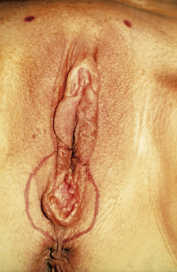
Patients generally present with a vulvar lump, although long-standing pruritus is common. The lesions may be raised, ulcerated, pigmented, or warty in appearance, and definitive diagnosis requires biopsy under local anesthesia. Most lesions occur on the labia majora; the labia minora are the next most common sites. Less commonly, the clitoris or the perineum is involved (Figure 40-2). About 5% of cases are multifocal.
Methods of Spread
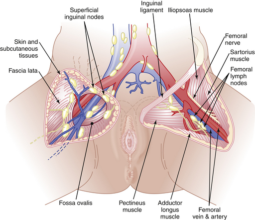
Vulvar cancer spreads by direct extension to adjacent structures, such as the vagina, urethra, and anus; by lymphatic embolization to regional lymph nodes; and by hematogenous spread to distant sites, including the lungs, liver, and bone. In most cases, the initial lymphatic metastases are to the inguinal lymph nodes, located between Camper’s fascia and the fascia lata. From these superficial nodes, spread occurs to the femoral nodes located medial to the femoral vein. Cloquet’s node, which is situated beneath the inguinal ligament, is the most cephalad of the femoral node group. From the inguinofemoral nodes, spread occurs to the pelvic nodes, particularly the external iliac group (Figure 40-3).

FIGURE 40-3 Lymphatic drainage of the vulva. Inguinal nodes are displayed on the right side of the groin, and femoral nodes are seen on the left side of the groin.
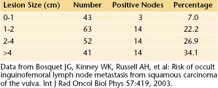
The incidence of lymph node metastases in vulvar cancer is about 30%. It is related to the size of the lesion (Table 40-1). About 5% of patients have metastases to pelvic lymph nodes. Such patients usually have three or more positive unilateral inguinofemoral lymph nodes. Hematogenous spread usually occurs late in the disease and rarely occurs in the absence of lymphatic metastases.
Staging
In 1989, the International Federation of Gynecology and Obstetrics (FIGO) Cancer Committee introduced a surgical staging system for vulvar cancer. This system was revised in 1994, and the present FIGO staging system is shown in Table 40-2.
TABLE 40-2 INTERNATIONAL FEDERATION OF GYNECOLOGY AND OBSTETRICS (FIGO) STAGING OF VULVAR CARCINOMA (1994)

Stay updated, free articles. Join our Telegram channel

Full access? Get Clinical Tree


Stage 0 | Carcinoma in situ, intraepithelial carcinoma |
Stage I | Tumor confined to the vulva or perineum, or both, and 2 cm or less in greatest dimension; no nodal metastasis |
Stage Ia | As above with stromal invasion ≤1 mm |
Stage Ib | As above with stromal invasion >1 mm |
Stage II | Tumor confined to the vulva or perineum, or both, and more than 2 cm in greatest dimension; no nodal metastasis |