97 Viral Infections
Epstein-Barr Virus
Epstein-Barr virus (EBV) is a common virus: most people become infected sometime in their lives. The clinical syndrome frequently associated with EBV is infectious mononucleosis, or “mono.” In socioeconomically disadvantaged areas, infants and children are most commonly affected, but adolescents are more commonly affected in affluent areas.
Etiology and Pathogenesis
EBV is transmitted by oral secretions and sexual intercourse, thus requiring close contact for transmission. After infection, the virus is excreted for many months and then intermittently for life. The incubation period is usually 30 to 50 days. The virus most likely spreads from the epithelial cells of the buccal mucosa to B-lymphocytes and then disseminates to the entire lymphoreticular system, including the liver and spleen. Similar to other herpesviruses, EBV stays latent in the body for life. EBV has been associated with a spectrum of proliferative disorders such as hemophagocytic syndrome, nasopharyngeal carcinoma, Burkitt lymphoma, and lymphoproliferative disorders.
Clinical Presentation
EBV infection causes a wide spectrum of clinical diseases. In many infants and young children, the symptoms are mild or unrecognized. The symptoms of infectious mononucleosis are malaise, fatigue, prolonged fever, sore throat, headache, nausea, and abdominal pain. Patients often have pharyngitis with exudates, lymphadenopathy, hepatomegaly, or splenomegaly. The incidence of dermatitis may be as high as 15% and is even more pronounced in children who were treated with ampicillin or amoxicillin; it is often called “ampicillin rash” (Figure 97-1). Rare complications of mononucleosis include airway obstruction, central nervous system (CNS) disorders, splenic rupture, thrombocytopenia, and hemolytic anemia.
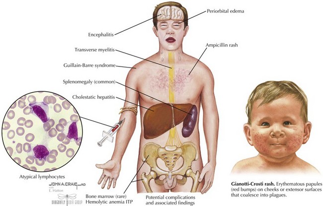
EBV is also associated with Gianotti-Crosti syndrome in which a symmetric rash is seen on the cheeks, extensor surfaces, or buttocks with multiple erythematous papules, which may coalesce into plaques. This may persist for 15 to 50 days and may sometimes look like atopic dermatitis (see Figure 97-1).
Differential Diagnosis
Several other pathogens may cause a mononucleosis-like illness, including cytomegalovirus (CMV), adenovirus, hepatitis viruses, HIV, rubella, and Toxoplasma gondii. Another infection that causes a similar clinical picture is streptococcal pharyngitis; however, it usually is not associated with hepatosplenomegaly.
Evaluation and Management
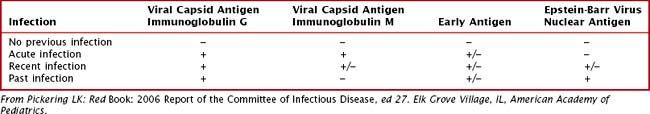
In 90% of cases, there is leukocytosis of 10,000 to 20,000 cells/mm3 with a predominance of lymphocytes with 20% to 40% atypical lymphocytes. There may be a mild thrombocytopenia but usually no purpura, and mild elevations of transaminases can occur. A nonspecific test for heterophile antibodies (also called Monospot) can be done. These are immunoglobulin M (IgM) antibodies that agglutinate sheep or horse red blood cells (RBCs) and usually appear during the first 2 weeks of illness and gradually disappear over a 6-month period. This test result is often negative in children younger than 4 years old. EBV can also be detected by serologic antibody testing. In the acute phase, there is a rapid immunoglobulin M (IgM) and IgG response to viral capsid antigen (VCA) and IgG to early antigen(EA). Positive Epstein-Barr virus nuclear antigen (EBNA) (nuclear antigen) antibody indicates past infection because this is not usually present until several weeks to months after infection (Table 97-1).
There is no specific treatment for infectious mononucleosis. Patients should not participate in contact sports until they have recovered or until the spleen normalizes. In severe cases with complications, a short course of corticosteroids may be helpful; however, there are no controlled data showing efficacy.
Future Directions
Further studies need to be done regarding the safety and efficacy of corticosteroids for treating complicated EBV infections. Research regarding treatment of EBV infections in immunocompromised patients is also needed. Investigations into associations between EBV, malignancies, and lymphoproliferative disorders will help elucidate these disease processes.
Measles
In the United States, the current rate of measles infection is less than one case per million people; however, historically, more than 90% of children were infected before the age of 15 years. This change is entirely attributable to the measles vaccine that was introduced in 1963. An outbreak that occurred between 1989 and 1991 resulted in 55,000 cases and prompted implementation of the two-dose vaccine. The majority of cases of measles are imported into the United States from abroad or are import related.
Etiology and Pathogenesis
Measles is transmitted by direct contact with large droplets or small droplet aerosols that enter through the respiratory tract or conjunctivae. Patients are contagious 3 days before the rash and up to 4 to 6 days afterward. The incubation period is 8 to 12 days. It is very contagious and affects 90% of exposed individuals. The virus causes necrosis of epithelium and a small vessel vasculitis of skin and oral mucosa. Infected cells fuse and cause multinucleated giant cells called Warthin-Finkeldey giant cells that are pathognomonic for measles.
Clinical Presentation
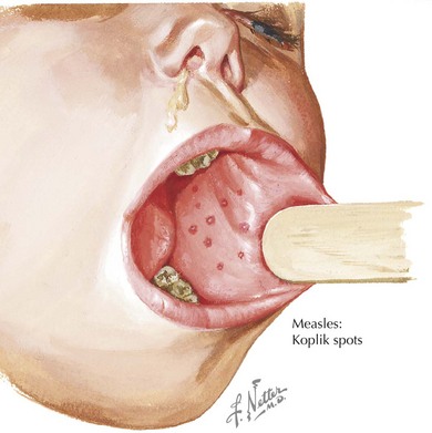
There are four phases to the infection: incubation period, prodrome, exanthem, and recovery. The prodrome period begins with a mild fever that increases to 103° to 105°F, conjunctivitis, coryza, and cough. Koplik spots appear 1 to 4 days before the rash. They appear as red lesions with a bluish white spot in the center and are usually on the inner aspects of the cheek. Rash usually appears after 2 to 4 days and begins on the face as discrete erythematous patches and spreads downward often becoming confluent (Figure 97-2). Lesions are also seen on the palms and soles. Rash fades in about 7 days and may leave desquamation of the skin. Cough can last up to 10 days.
Complications include otitis media, pneumonia, laryngotracheobronchitis, seizures, and diarrhea. In rare cases, measles may cause acute encephalitis and subacute sclerosing panencephalitis (SSPE), a degeneration of the CNS usually occurring an average of 7 years later.
Differential Diagnosis
Measles may sometimes be confused with other viruses that cause exanthems such as rubella, adenovirus, enterovirus, EBV, human herpesvirus-6 (HHV-6), and parvovirus. Mycoplasma pneumoniae and group A streptococci (GAS) can also produce similar rashes. Several clinical findings are similar in Kawasaki’s disease, but there is a more prominent cough and no thrombocytosis in measles. Drug reactions should also be in the differential diagnosis.
Evaluation and Management
The diagnosis of measles is usually clinical; however, laboratory findings can include decreased white blood cell count, and a normal erythrocyte sedimentation rate and C-reactive protein. IgM antibody can be detected 1 to 2 days after the onset of rash and remains for 1 month and is therefore indicative of acute infection. A fourfold increase in IgG antibodies after 2 to 4 weeks can also be diagnostic. There is no specific treatment for measles; however, low vitamin A levels are associated with increased morbidity and mortality. Therefore, vitamin A supplementation is recommended. No antibiotic prophylaxis is recommended.
Prevention
Vaccination is the best form of prevention. The first dose is recommended between 12 and 15 months followed by a second dose at 4 to 6 years of age. Because the vaccine is a live-attenuated vaccine, it should not be given to pregnant women or severely immunocompromised children. The MMR (mumps, measles, and rubella) vaccine can cause fever (6-12 days after), rash (7-12 days after) and rarely a transient thrombocytopenia. Fortunately, several large and well-designed scientific studies have convincingly shown that there is no evidence that the measles vaccine causes autism.
Postexposure prophylaxis can be administered via vaccine if it is less than 72 hours after exposure or immune globulin up to 6 days after exposure.
Future Directions
Continued educational efforts need to be undertaken to assure parents about the necessity and the safety of the measles vaccine. Preventing measles from becoming endemic again in the United States will require maintenance of a high level of immunity through vaccination. Eliminating measles from developing countries through vaccines remains a continuing effort.
Herpes Simplex Virus
There are 2 types of herpes simplex virus (HSV), type 1 and type 2, that can cause a variety of illnesses depending on the host and the site of infection. A primary herpes infection occurs in those who have never been infected with either HSV-1 or HSV-2. A nonprimary first infection occurs when an individual who was previously infected with one type of HSV then becomes infected with another type. A recurrent infection is a reactivation of the virus from the latent state. HSV can also cause severe neonatal infection (see Chapter 105).
Etiology and Pathogenesis
HSV is ubiquitous and is transmitted through direct contact with infected mucocutaneous surfaces. Even if a person is asymptomatic, the virus can be intermittently shed; however, the greatest concentration of virus is shed during symptomatic primary infections. Although HSV-1 is thought to infect oral mucosa and HSV-2 is thought to cause genital infections, either type can cause initial infections in any mucosa surface. However, HSV-1 is more likely to cause recurrent oral infections, and HSV-2 is more likely to cause recurrent genital infections.
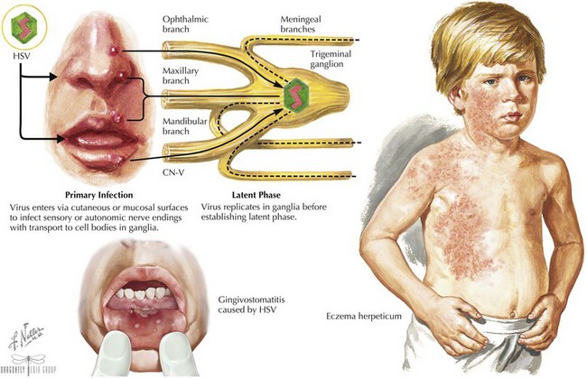
The virus enters through mucosal surfaces and then spreads via nerve endings to sensory ganglia. Some viruses then establish latency in these sensory neurons (Figure 97-3). The incubation period is usually 2 days to 2 weeks.
Clinical Presentation
The most common clinical manifestation of primary infection in children is gingivostomatitis and is usually caused by HSV-1. It causes sudden onset of pain in the mouth often manifested as refusal to eat, drooling, and high fevers. The gums become very swollen, and vesicles that are usually grouped on an erythematous base are seen throughout the oral cavity, including the gums, lips, tongue, palate, tonsils, and pharynx. The vesicles can progress to ulcers, and lymphadenopathy is often seen (see Figure 97-3). The illness usually resolves in 7 to 14 days.
HSV infections can occur on any skin surface that may have breakdown or trauma. Herpetic whitlow is a term used for HSV infections of the fingers or toes. Lesions and pain usually last for 10 days, and complete recovery usually occurs in 18 to 20 days. Eczema herpeticum is described in patients with a history of eczema who are superinfected with HSV (see Figure 97-3). In addition to severe rash, patients can present with high fevers, malaise, and lymphadenopathy. Other areas of the body that may be affected by HSV include the conjunctiva, cornea, and retina as well as the CNS, causing encephalitis and aseptic meningitis. Thus, any vesicles around the eyes should prompt an ophthalmologic examination. HSV has also been implicated in erythema multiforme and Bell’s palsy.

Stay updated, free articles. Join our Telegram channel

Full access? Get Clinical Tree

