Chapter 350 Viral Hepatitis
Viral hepatitis continues to be is a major health problem in both developing and developed countries. This disorder is caused by at least 5 pathogenic hepatotropic viruses recognized to date: hepatitis A, B, C, D, and E viruses (Table 350-1). Many other viruses (and diseases) can cause hepatitis, usually as 1 component of a multisystem disease. These include herpes simplex virus (HSV), cytomegalovirus (CMV), Epstein-Barr virus (EBV), varicella-zoster virus, HIV, rubella, adenoviruses, enteroviruses, parvovirus B19, and arboviruses (Table 350-2).
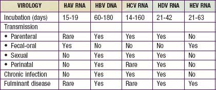
Table 350-2 CAUSES AND DIFFERENTIAL DIAGNOSIS OF HEPATITIS IN CHILDREN
INFECTIOUS
NON-VIRAL LIVER INFECTIONS
AUTOIMMUNE
METABOLIC
TOXIC
ANATOMIC
HEMODYNAMIC
NON-ALCOHOLIC FATTY LIVER DISEASE
From Wyllie R, Hyams JS, editors: Pediatric gastrointestinal and liver disease, ed 3, Philadelphia, 2006, Saunders.
The hepatotropic viruses are a heterogeneous group of infectious agents that cause similar acute clinical illness. In most pediatric patients, the acute phase causes no or mild clinical disease. Morbidity is related to rare cases of acute liver failure (ALF) triggered in susceptible patients and to the chronic disease state and attendant complications that three of these viruses (hepatides B, C, and D) can cause.
Issues Common to All Forms of Viral Hepatitis
Differential Diagnosis
Often what brings the patient with hepatitis to medical attention is clinical icterus, which is a mixed or conjugated (direct) reacting hyperbilirubinemia.
Symptomatic infection results in icteric skin and mucous membranes. The liver is usually enlarged and tender to palpation and percussion. Splenomegaly and lymphadenopathy can be present. Extrahepatic symptoms are more readily seen in HBV and HCV infections (rashes, arthritis). Clinical signs of altered sensorium and hyperreflexivity should be carefully sought, because they mark the onset of encephalopathy and ALF.
The differential diagnosis varies with age of presentation.
In the newborn period, infection is a common cause of conjugated hyperbilirubinemia; the infectious cause is either a bacterial agent (Escherichia coli, Listeria, Syphilis) or one of the nonhepatotropic viruses (HSV, enteroviruses, CMV). Metabolic and anatomic causes (tyrosinemia, biliary atresia, genetic forms of intrahepatic cholestasis, and choledochal cysts) should always be excluded.
In later childhood, extrahepatic obstruction (gallstones, primary sclerosing cholangitis, pancreatic pathology), inflammatory conditions (autoimmune hepatitis, juvenile rheumatoid arthritis, Kawasaki disease, immune dysregulation), infiltrative disorders (malignancies), toxins and medications, metabolic disorders (Wilson disease, cystic fibrosis), and infection (EBV, varicella, malaria, leptospirosis, syphilis) should be ruled out.
Pathogenesis
The acute response of the liver to hepatotropic viruses involves a direct cytopathic and an immune-mediated injury. The entire liver is involved. Necrosis is usually most marked in the centrilobular areas. An acute mixed inflammatory infiltrate predominates in the portal areas but also affects the lobules. The lobular architecture remains intact, although balloon degeneration and necrosis of single or groups of parenchymal cells occurs commonly. Fatty change is rare except with HCV infection. Bile duct proliferation but not bile duct damage is common. Diffuse Kupffer cell hyperplasia is noticeable in the sinusoids. Neonates often respond to hepatic injury by forming giant cells.
In fulminant hepatitis, parenchymal collapse occurs on the just-described background.
With recovery, the liver morphology returns to normal within 3 mo of the acute infection. If chronic hepatitis develops, the inflammatory infiltrate settles in the periportal areas and often leads to progressive scarring. Both of these hallmarks of chronic hepatitis are seen in cases of HBV and HCV.
Common Biochemical Profiles in the Acute Infectious Phase
Acute liver injury caused by the hepatotropic viruses manifests in 3 main functional liver biochemical profiles. These serve as an important guide to supportive care and monitoring in the acute phase of the infection for all viruses.
As a reflection of cytopathic injury to the hepatocytes, there is a rise in serum levels of alanine aminotransferase (ALT) and aspartate aminotransferase (AST). The magnitude of enzyme elevation does not correlate with the extent of hepatocellular necrosis and has little prognostic value. There is usually slow improvement over several weeks, but AST and ALT levels lag behind the serum bilirubin level, which tends to normalize first. Rapidly falling aminotransferase levels can predict a poor outcome, particularly if their decline occurs in conjunction with a rising bilirubin level and a prolonged prothrombin time; this combination of findings usually indicates that massive hepatic injury has occurred.
Cholestasis, defined by elevated serum conjugated bilirubin levels, results from abnormal bile flow at the canalicular and cellular level due to hepatocyte damage and inflammatory mediators. Elevation of serum alkaline phosphatase (ALP), 5′-nucleotidase, γ-glutamyl transpeptidase (GGT), and urobilinogen all mark cholestasis. Improvement tends to parallel the acute hepatitis phase. Absence of cholestatic markers does not rule out progression to chronicity in HCV or HBV infections.
The most important marker of liver injury is altered synthetic function. Monitoring of synthetic function should be the main focus in clinical follow-up to define the severity of the disease. In the acute phase, the degree of liver synthetic dysfunction guides treatment and helps to establish intervention criteria. Abnormal liver synthetic function is a marker of liver failure and is an indication for prompt referral to a transplant center. Serial assessment is necessary because liver dysfunction does not progress linearly. Synthetic dysfunction is reflected by a combination of abnormal protein synthesis (prolonged prothrombin time, high international normalized ratio [INR], low serum albumin levels), metabolic disturbances (hypoglycemia, lactic acidosis, hyperammonemia), poor clearance of medications dependent on liver function, and altered sensorium with increased deep tendon reflexes (hepatic encephalopathy; Chapter 356).
Hepatitis A
Hepatitis A virus (HAV) infection is the most prevalent of the 5. This virus is also responsible for most forms of acute and benign hepatitis; although fulminant hepatic failure can occur, it is rare and occurs more often in adults than in children.
Etiology
HAV is an RNA virus, a member of the picornavirus family. It is heat stable and has limited host range—namely, the human and other primates.
Epidemiology
HAV infection occurs throughout the world but is most prevalent in developing countries. In the United States, 30-40% of the adult population has evidence of previous HAV infection. Hepatitis A is thought to account for ∼50% of all clinically apparent acute viral hepatitis in the United States. As a result of aggressive implementation of a childhood vaccination strategy, the prevalence of symptomatic HAV cases in the United States has declined significantly. However, outbreaks in daycare centers (where the spread from young, nonicteric, infected children can occur easily) as well as multiple foodborne and waterborne outbreaks have justified the implementation of a universal vaccination program.
HAV is highly contagious. Transmission is almost always by person-to-person contact through the fecal-oral route. Perinatal transmission occurs rarely. No other form of transmission is recognized. HAV infection during pregnancy or at the time of delivery does not appear to result in increased complications of pregnancy or clinical disease in the newborn. In the USA, increased risk of infection is found in contacts with infected persons, child-care centers, and household contacts. Infection has also been associated with contact with contaminated food or water and after travel to endemic areas. Common source foodborne and waterborne outbreaks have occurred, including several due to contaminated shellfish, frozen berries, and raw vegetables; no known source is found in about half of the cases. The mean incubation period for HAV is ∼3 wk. Fecal excretion of the virus starts late in the incubation period, reaches its peak just before the onset of symptoms, and resolves by 2 wk after the onset of jaundice in older subjects. The duration of viral excretion is prolonged in infants. The patient is therefore contagious before clinical symptoms are apparent and remains so until viral shedding ceases.
Clinical Manifestations
HAV is responsible for acute hepatitis only. Often, this is an anicteric illness, with clinical symptoms indistinguishable from other forms of viral gastroenteritis, particularly in young children.
The illness is much more likely to be symptomatic in older adolescents or adults, in patients with underlying liver disorders, and in those who are immunocompromised. It is characteristically an acute febrile illness with an abrupt onset of anorexia, nausea, malaise, vomiting, and jaundice. The typical duration of illness is 7-14 days (Fig. 350-1).

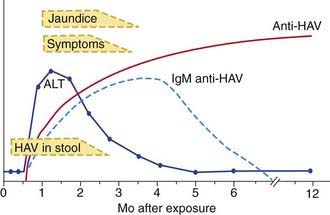
Figure 350-1 The serologic course of acute hepatitis A. ALT, alanine aminotransferase; HAV, hepatitis A virus.
(From Goldman L, Ausiello D: Cecil textbook of medicine, ed 22, Philadelphia, 2004, Saunders, p 913.)
Other organ systems can be affected during acute HAV infection. Regional lymph nodes and the spleen may be enlarged. The bone marrow may be moderately hypoplastic, and aplastic anemia has been reported. Tissue in the small intestine might show changes in villous structure, and ulceration of the gastrointestinal tract can occur, especially in fatal cases. Acute pancreatitis and myocarditis have been reported, though rarely, and nephritis, arthritis, vasculitis, and cryoglobulinemia can result from circulating immune complexes.
Diagnosis
Acute HAV infection is diagnosed by detecting antibodies to HAV, specifically, anti-HAV (IgM) by radioimmunoassay or, rarely, by identifying viral particles in stool. A viral polymerase chain reaction (PCR) assay is available for research use (Table 350-3). Anti-HAV is detectable when the symptoms are clinically apparent, and it remains positive for 4-6 mo after the acute infection. A neutralizing anti-HAV (IgG) is usually detected within 8 wk of symptom onset and is measured as part of a total anti-HAV in the serum. Anti-HAV (IgG) confers long-term protection.
Rises in serum levels of ALT, AST, bilirubin, ALP, 5′-nucleotidase, and GGT are almost universally found and do not help to differentiate the cause of hepatitis.
Complications
Although most patients achieve full recovery, two distinct complications can occur.
ALF from HAV infection is a rare but not infrequent complication of HAV. Those at risk for this complication are adolescents and adults, but also patients with underlying liver disorders or those who are immunocompromised. The height of HAV viremia may be linked to the severity of hepatitis. Whereas in the United States, HAV represents <0.5% of pediatric-aged ALF, it is responsible for up to 3% mortality in the adult population with ALF. In endemic areas of the world, HAV constitutes up to 40% of all cases of pediatric ALF.
HAV can progress to a prolonged cholestatic syndrome that waxes and wanes over several months. Pruritus and fat malabsorption are problematic and require symptomatic support with antipruritic medications and fat-soluble vitamins. This syndrome occurs in the absence of any liver synthetic dysfunction and resolves with no sequelae.
Treatment
There is no specific treatment for hepatitis A. Supportive treatment consists of intravenous hydration as needed and antipruritic agents and fat-soluble vitamins for the prolonged cholestatic form of disease. Serial monitoring for signs of ALF and, if ALF is diagnosed, a prompt referral to a transplantation center can be lifesaving.
Prevention
Patients infected with HAV are contagious for 2 wk before and ~7 days after the onset of jaundice and should be excluded from school, child care, or work during this period. Careful handwashing is necessary, particularly after changing diapers and before preparing or serving food. In hospital settings, contact and standard precautions are recommended for 1 wk after onset of symptoms.
Immunoglobulin
Indications for intramuscular administration of immunoglobulin (IG) (0.02 mL/kg) include pre-exposure and postexposure prophylaxis (Table 350-4).
Table 350-4 HEPATITIS A VIRUS PROPHYLAXIS
PRE-EXPOSURE PROPHYLAXIS (TRAVELERS TO ENDEMIC REGIONS) | ||
---|---|---|
Age | Exposure | Dose |
<1 yr of age | Expected <3 mo | Ig 0.02 mL/kg |
Expected 3-5 mo | Ig 0.06 mL/kg | |
Expected long term | Ig 0.06 mL/kg at departure and every 5 mo thereafter | |
≥1 yr of age | Healthy host | HAV vaccine |
Immunocompromised host, or one with chronic liver disease or chronic health problems | HAV vaccine and Ig 0.02 mL/kg |
POSTEXPOSURE PROPHYLAXIS* | |
---|---|
Exposure | Recommendations |
≤2 wk since exposure | < 1 y of age: IG 0.02 mL/kgImmunocompromised host, or host with chronic liver disease or chronic health problems: IG 0.02 mL/kg and HAV vaccine>1 yr and healthy host: HAV vaccine, IG remains optionalSporadic non-household or close contact exposure: prophylaxis not indicated* |
>2 wk since exposure | None |
Ig, immunoglobulin.
* Decision for prophylaxis in non-household contacts should be tailored to individual exposure and risk.
IG is recommended for pre-exposure prophylaxis for susceptible travelers to countries where HAV is endemic, and it provides effective protection for up to 3 mo. HAV vaccine given any time before travel is preferred for pre-exposure prophylaxis in healthy persons, but IG ensures an appropriate prophylaxis in children <1 yr of age, patients allergic to a vaccine component, or those who elect not to receive the vaccine. If travel is planned in <2 wk, older patients, immunocompromised hosts, and those with chronic liver disease or other medical conditions should receive both IG and the HAV vaccine.
IG as prophylaxis in postexposure situations should be used as soon as possible (not effective >2 wk after exposure). It is exclusively used for children <12 mo of age, immunocompromised hosts, those with chronic liver disease or in whom vaccine is contraindicated; IG is preferably used in patients > 40 yr of age. IG is optional in healthy persons 12 mo-40 yr, in whom HAV vaccine is preferred. An alternative approach is to immunize previously unvaccinated patients who are ≥12 mo at the age-appropriate vaccine dosage as soon as possible. IG is not routinely recommended for sporadic nonhousehold exposure (e.g., protection of hospital personnel or schoolmates).
Vaccine
The availability of two inactivated, highly immunogenic, and safe HAV vaccines has had a major impact on the prevention of HAV infection. Both vaccines are approved for children >1 yr of age. They are administered intramuscularly in a 2-dose schedule, with the 2nd dose given 6-12 mo after the 1st dose. Seroconversion rates in children exceed 90% after an initial dose and approach 100% after the 2nd dose; protective antibody titer persists at least for 10 yr. The immune response in immunocompromised persons, older patients, and those with chronic illnesses may be suboptimal; in those patients, combining the vaccine with IG for pre- and postexposure prophylaxis is indicated. HAV vaccine may be administered simultaneously with other vaccines. A combination HAV and HBV vaccine is currently undergoing trials in adults. For healthy persons >1 yr of age, vaccine is preferable to immunoglobulin for pre-exposure and postexposure prophylaxis (see Table 350-3).
Hepatitis B
Etiology
HBV is a member of the Hepadnaviridae family. HBV has a circular, partially double-stranded DNA genome composed of ∼3,200 nucleotides. Four genes have been identified: the S (surface), C (core), X, and P (polymer) genes. The surface of the virus includes particles designated hepatitis B surface antigen (HBsAg), which is a 22 nm diameter spherical particle and a 22 nm wide tubular particle with a variable length of up to 200 nm. The inner portion of the virion contains hepatitis B core antigen (HBcAg), the nucleocapsid that encodes the viral DNA, and a nonstructural antigen called hepatitis B e antigen (HBeAg), a nonparticulate soluble antigen derived from HBcAg by proteolytic self-cleavage. HBeAg serves as a marker of active viral replication and usually correlates with HBV DNA levels. Replication of HBV occurs predominantly in the liver but also occurs in the lymphocytes, spleen, kidney, and pancreas.

Full access? Get Clinical Tree

