28 Vasculitis
Vasculitis is inflammation of a blood vessel, which can occur as a primary process or may be secondary to another disease. For example, rheumatic illnesses, such as systemic lupus erythematosus (SLE) or juvenile dermatomyositis, may have vasculitis as a secondary process. Overall, the childhood vasculitides are a rare group of disorders. The occurrence of vasculitis in patients younger than 17 years old is approximately 20 per 100,000 children. However, specific diseases, such as Churg-Strauss syndrome, can be even rarer. In addition, the prevalence of diseases may be different based on the population studied. For example, Behçet’s disease occurs more frequently in the Turkish or Japanese populations (i.e., origins along the “Silk Road”).
This chapter focuses on the primary vasculitides. A general overview is presented followed by a more detailed discussion of Henoch-Schönlein purpura (HSP) and Kawasaki disease (KD), the two most common childhood vasculitides.
Etiology and Pathogenesis
The underlying cause of vasculitis has yet to be elucidated; however, similar to many chronic diseases, there is likely a complex interplay of genetic and environmental factors. Some theories suggest that the inflammation is attributable to the involvement of humoral factors, as seen in the antineutrophil cytoplasmic antibodies (ANCA)–associated vasculitides. Others suggest abnormal regulation of immune complex formation is contributory, as in HSP. Lymphocyte involvement has also been implicated, specifically T-regulatory cell dysfunction. Additionally, antecedent infections, particularly streptococcal infections, have been suggested as a cause in many of the vasculitides such as HSP, Wegener’s granulomatosis, and polyarteritis nodosa (PAN).
Classification
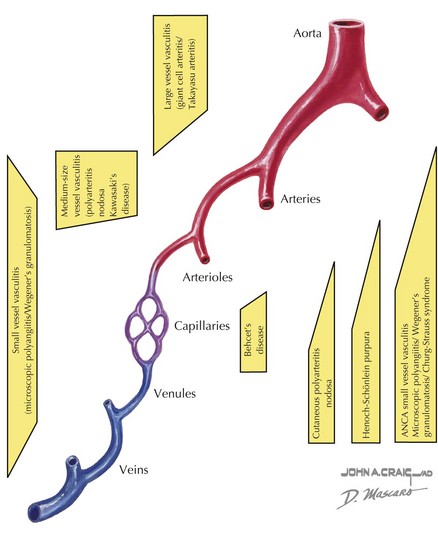
Vasculitis can be classified based on the involvement of primarily large, medium, or small vessels (Figure 28-1). In addition, certain vasculitides have a predilection for arteries, veins, or both. The vasculitides can be further classified as granulomatous or nongranulomatous. The granulomatous diseases include Takayasu arteritis, Wegener’s granulomatosis, and Churg-Strauss syndrome (see Chapter 30). The nongranulomatous vasculitides include PAN, KD, microscopic polyangiitis, HSP, cutaneous leukocytoclastic vasculitis, and essential cryoglobulinemic vasculitis.
Clinical Presentation
Vasculitis often presents with vague symptoms and multiorgan involvement. Nonspecific systemic features may include prolonged fever without any clear source, hypertension, fatigue, malaise, weight loss, and rash. As a consequence, the diagnosis is often delayed and requires a high index of suspicion. Definitive diagnosis often relies on imaging (angiography) and tissue biopsy. The history should include questions about recent infections, medication exposures, and a detailed family history.
Certain patterns of clinical symptoms and organ involvement may be suggestive of a specific vasculitis (Box 28-1). For example, whereas palpable purpura, arthralgias, abdominal pain, and renal disease suggest HSP, persistent fever, conjunctivitis, cervical lymphadenopathy, extremity swelling, mucocutaneous changes, and rash suggest KD (see below). Microscopic polyangiitis is associated with high titers of pANCA (protoplasmic-staining antineutrophil cytoplasmic antibodies) and affects mainly the pulmonary and renal systems. Hypersensitivity vasculitis is a necrotizing vasculitis that presents with a papular rash that may be red or blistering and is often associated with infection or medication exposure. Hypocomplementemic urticarial vasculitis presents with urticarial skin lesions and low serum complement levels of C4 and C3. Behçet’s disease is a unique systemic vasculitis that affects both arteries and veins. The classic triad of Behçet’s disease includes oral ulcers, uveitis, and genital ulcers; however, any organ system can be affected.
PAN is a necrotizing vasculitis affecting the medium-sized arteries and can present with systemic disease or in a limited form that only involves the skin and joints. It occurs in school-aged children, and there is typically a history of a preceding upper respiratory infection or streptococcal pharyngitis. In unvaccinated children, hepatitis B can be causative. Symptoms include prolonged fevers, malaise, calf pain, testicular pain, and weight loss. Physical examination findings include painful nodules (particularly on the feet), livedo reticularis (see Figure 29-2), myalgias, and arthritis of large joints. In the systemic form, any organ system can be affected; thus, hypertension, renal abnormalities, gastrointestinal involvement, and coronary disease can be seen. Of the large vessel diseases, temporal arteritis is not seen in childhood. Takayasu arteritis, the third most common childhood vasculitis, preferentially affects the large branches of the aorta. Examination may reveal bruits, hypertension, and absent pulses.
Physical Examination
Hypertension is common with many of the vasculitides. Furthermore, Takayasu arteritis classically presents with a blood pressure difference of greater than 10 mm Hg between arms. Thus, the physical examination should include evaluation of blood pressure in all four extremities. Careful auscultation for bruits (carotid, aortic, and abdominal vessels) and palpation of all peripheral pulses is vital; absent peripheral pulses (claudication) may help narrow the differential diagnosis and areas of vessel involvement. A thorough skin examination is warranted, since the presence of nodules, vasculitis lesions, ulcerations, microinfarctions, or livedo reticularis may help aid in diagnosis. A neurological examination should focus on evidence of neuropathy; some of the vasculitides, such as PAN, are associated with mononeuritis multiplex. Two specific areas where blood vessels can be directly visualized are the ocular examination and the nailfold capillaries. As disease progresses, more specific signs may develop, but morbidity also increases. Therefore, a high clinical suspicion for vasculitis is important.

Stay updated, free articles. Join our Telegram channel

Full access? Get Clinical Tree

