123 Vascular Disorders
Vascular lesions of the skin are a common pediatric problem with a wide range of clinical presentations. In 1996, the International Society for the Study of Vascular Anomalies adopted Mulliken and Glowacki’s classification (Table 123-1). There are two categories based on biologic and clinical characteristics: vascular tumors and vascular malformations. Vascular tumors are neoplasms of vascular structures that grow by hyperplasia. Vascular malformations are local anomalies in vascular development that do not demonstrate proliferation.
Table 123-1 International Society for the Study of Vascular Anomalies Classification of Vascular Anomalies
Vascular Tumors | Vascular Malformations |
---|---|
VM, venous malformation.
Adapted from ISSVA classification as reported in Color Atlas of Vascular Tumors & Vascular Malformations.
Vascular Tumors
Recent classification distinguishes three major types of vascular tumors, hemangiomas, tufted angiomas, and hemangioendotheliomas. Although these vascular tumors are generally benign, they have significant associated clinical consequences.
Infantile Hemangiomas
Etiology and Pathogenesis
Hemangiomas of infancy are the most common infantile tumors, occurring in up to 1% to 2% of all infants and up to 10% in whites. These benign proliferations of endothelial cells affect females four times more often than males. The growing incidence of hemangiomas has been attributed to an association between hemangiomas and premature birth. Other factors that are associated with an increased incidence of hemangiomas include multiple gestations, assisted reproduction, chorionic villus sampling, increased maternal age, and preeclampsia. However, many of these factors may not be independent. Although most hemangiomas are sporadic occurrences, a positive family history does increase the incidence, and both autosomal dominant inheritance and syndromic associations are reported.
Clinical Manifestations
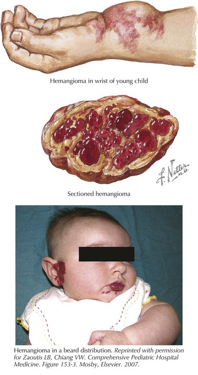
Hemangiomas are classified as superficial, deep, or mixed lesions. Superficial hemangiomas, involving the papillary dermis, are red and protuberant. Superficial hemangiomas have well-defined borders. Deep lesions invade the reticular dermis and superficial fat. They tend to have a blue-purple discoloration with normal skin texture. The margins of deep lesions may be ill defined. Most lesions have both superficial and deep components (Figure 123-1). A mature hemangioma contains characteristics of capillaries, venules, and arterioles.
Although subtle skin changes may be present at birth, most hemangiomas do not become evident until the first few weeks of life when they undergo a period of rapid growth that may last 6 to 9 months. Growth then slows, and most cease growing by 10 to 12 months of age. Spontaneous involution and regression occurs after the cessation of growth. The first sign of regression is the appearance of grayish changes on the surface of the hemangioma. Although the vascular elements of the tumor may completely regress, they are often replaced by fibrofatty tissue that results in permanent skin changes (30%), including scarring, atrophy, telangiectasias, or permanent discoloration. Although some lesions will not fully regress, more than 60% of lesions have completed their regression by age 5 years, with almost all lesions completed by age 9 years.
Management
Most infantile hemangiomas are of little clinical significance and require no therapy. Treatment is based primarily on the location, size, and potential for complications. Lesions that affect vision, breathing, eating, or bowel habits or those that ulcerate or are large and likely to lead to significant cosmetic abnormalities may necessitate therapy. The mainstays of treatment include pulsed-dye laser therapy and corticosteroids. Pulsed-dye laser results in a high response rate for superficial hemangiomas but is typically reserved for ulcerated lesions because superficial hemangiomas will likely regress fully anyway. Steroid-resistant, life-threatening lesions may require treatment with chemotherapeutic agents such as vincristine or interferon-α (INF-α). Recent data suggest the efficacy of oral propranolol in shortening the course of infantile hemangiomas. Although propranolol therapy is well tolerated, care should be taken in children with abnormal vasculature, reactive airway disease, or underlying cardiac conditions.
The most common complication of rapidly proliferating hemangiomas is ulceration. Ulceration is most common in perineal and perioral hemangiomas and can be quite painful. Ulcerated hemangiomas are also at risk for superinfection. Hemangiomas in select locations may have unique complications. Nasal tip hemangiomas cause significant disfigurement. Periocular hemangiomas disrupt visual development, and early referral to an ophthalmologist is prudent. Hemangiomas along the jaw line or neck (beard distribution) of the head and neck (see Figure 123-1) have a high association with airway lesions that may cause life-threatening airway obstruction. Spinal dysraphism is associated with midline lumbosacral lesions, and magnetic resonance imaging (MRI) is indicated. Infants with multiple cutaneous hemangiomas warrant evaluation for visceral hemangiomas.

Stay updated, free articles. Join our Telegram channel

Full access? Get Clinical Tree

