Urticaria and Angioedema

20 Urticaria and Angioedema


Urticaria, or hives, is a pruritic and episodic rash that most commonly occurs without an identifiable trigger. It occurs in approximately 25% of the population. The occurrence of urticaria increases to 50% in those affected by allergic disorders, such as asthma, allergic rhinitis, or atopic dermatitis. Acute urticaria, defined as symptoms lasting less than 6 weeks, accounts for two-thirds of all urticaria. Chronic urticaria, which can be subdivided into chronic idiopathic urticaria (CIU) and physical urticaria, is classified by symptom duration of greater than 6 weeks with symptoms on at least 2 days per week. CIU occurs in approximately 0.1% to 3% of the population and is more prevalent in females. Features of physical urticarias can be common, especially dermatographism, which occurs in almost half of the general population, including those with no history of chronic urticaria.


Chronic urticaria can have a significant social and financial impact. The hallmark of urticaria is pruritus, which contributes to difficulty sleeping and in activities of daily life. Urticaria can be a frustrating disease for patients because typically there is no identifiable trigger for exacerbations, leading to an unpredictable course. The effect on quality of life is comparable to that of coronary heart disease and atopic dermatitis. Patients with chronic urticaria also have multiple medications, medical visits, work and school absences, and use of the emergency department, all of which contribute to the economic burden of the disease.


Etiology and Pathogenesis


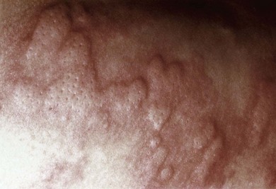
Urticaria is characterized by the waxing and waning appearance of pruritic, erythematous papules or plaques with superficial swelling of the dermis (Figure 20-1). The swelling observed with urticaria and angioedema results from the movement of plasma from small blood vessels into adjacent connective tissue. Unlike atopic dermatitis, the pruritus of urticaria is driven by histamine. Histologically, urticarial lesions consist of a lymphocyte-predominant perivascular infiltrate. In CIU, the cellular infiltrate is similar to that seen in allergen-induced late phase skin response, and the cytokine profile is both TH1 and TH2.


Chronic Idiopathic Urticaria


CIU, as the name implies, typically lacks an identifiable, consistent trigger. The episodic onset of disease is more commonly observed in adults than children. The disease duration is on average 3 to 5 years, with greater duration if severe disease, angioedema, or autoimmune features are present. The pathogenesis of CIU is still unknown, although autoantibodies and cells typically involved in IgE-mediated reactions, such as mast cells and basophils, have been implicated. About 30% to 40% of patients with CIU are classified as having autoimmune urticaria, a subgroup of CIU defined by the presence of histamine-releasing autoantibodies. The majority of these autoantibodies are directed against the α subunit of the high-affinity receptor, FcεRI; the remainder target IgE. These autoantibodies have been hypothesized to be pathogenic, although this remains controversial because they can also be found in healthy individuals, in addition to those with other autoimmune diseases, such as systemic lupus and dermatomyositis. Injection of autologous serum, also known as the autologous serum skin test (ASST), leads to a wheal-and-flare response in CIU patients, suggesting that the causative agent is in the serum. Patients with CIU have a higher prevalence of other autoantibodies such as antimicrosomal and antithyroglobulin thyroid autoantibodies and a higher frequency of certain HLA class II alleles (DR4, DQ8) associated with autoimmunity.


More recently, the role of mast cells and basophils has been investigated. Skin biopsies of patients with CIU demonstrate mast cell degranulation accompanied by increased mast cell releasibility that reverses with disease remission. Basophils, which typically are not present in the skin, are observed in both lesional and nonlesional CIU skin biopsies. Basopenia has been described in CIU and is a marker of more severe disease. More recently, basophil activation markers have been shown to be enhanced in CIU patients along with defects in basophil signaling through the IgE receptor.


Physical Urticaria


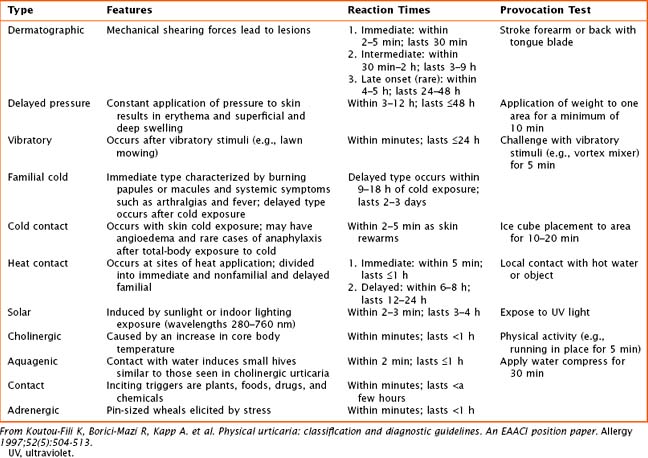
Physical urticarias have a specific trigger that directly induces hives within minutes, with hives lasting minutes to a few hours. Physical urticarias are further classified into multiple subtypes (Table 20-1), each having a different physical trigger; thus, an individual can have more than one subtype. Mast cell degranulation is included in the pathogenesis of most subtypes of physical urticaria, including dermatographic, cholinergic, cold, and solar urticaria, but serum immunoglobulin may also be involved as demonstrated by passive transfer experiments.


Angioedema


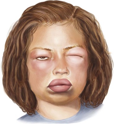
Angioedema, which is a swelling of the dermis, subcutaneous, and submucosal tissues, often coexists with urticaria but typically persists past 24 hours (Figure 20-2). Angioedema is described as painful or burning in quality. The lack of pruritus in angioedema may be caused by fewer mast cells in the lower dermis and subcutis. Idiopathic angioedema coexists with urticaria, occurring in up to 50% of those with CIU. Angiotensin-converting enzyme (ACE) inhibitors can also trigger angioedema via the bradykinin pathway.


Clinical Presentation


Urticarial lesions are intensely pruritic and can affect any location on the body. They are typically transient, lasting less than 24 hours. Unlike atopic dermatitis, excoriation is not a typical finding in urticaria, regardless of the degree of pruritus experienced. Lesions can vary in size and can be confluent. Similar to urticaria, angioedema can occur anywhere on the body, although it frequently involves face, lips, tongue, throat, and extremities, but unlike urticaria, it commonly involves mucous membranes. Cold urticaria is the exception because it may involve swelling of the tongue or palate. Urticarial lesions usually demonstrate complete resolution without skin pigment changes.


Patients with long-lasting lesions or the presence of other systemic symptoms should be evaluated for other diseases (see Box 20-1). Urticarial vasculitis is classified by painful or pruritic lesions lasting longer than 48 hours and may leave residual skin changes unrelated to excoriation. Concurrent systemic complaints or abnormal laboratory findings indicative of an inflammatory process, such as an elevated erythrocyte sedimentation rate (ESR) or C-reactive protein (CRP) or low complement levels, are seen. A skin biopsy is required to rule out urticarial vasculitis. Urticaria pigmentosa, a subset of mastocytosis, may mimic urticaria, although these lesions are typically pigmented and last longer than urticaria. The presence of fever with urticaria can occur in Schnitzler’s syndrome and Muckle-Wells syndrome. Schnitzler’s syndrome is described as recurrent urticaria with arthralgia, fever, and elevation in inflammatory markers in association with an elevation in IgM. Muckle-Wells syndrome is a periodic fever syndrome with urticaria associated with periodic, unexplained fevers.


Jun 19, 2016 | Posted by in PEDIATRICS | Comments Off on Urticaria and Angioedema

Full access? Get Clinical Tree

Get Clinical Tree app for offline access