Indications for urodynamic testing
Complicated history
Stress incontinence before surgical correction
Urge incontinence not responsive to therapy
Recurrent urinary loss after previous surgery for stress incontinence
Frequency, urgency, and pain syndromes not responsive to therapy
Nocturnal enuresis not responsive to therapy
Lower urinary tract dysfunction after pelvic radiation or radical pelvic surgery
Neurologic disorders
Continuous leakage
Suspected voiding difficulties
The most recent report by International Consultation on Incontinence (ICI) states that the role of urodynamic studies in clinical practice should be (1) to identify or to rule out factors contributing to the LUT dysfunction and assess their relative importance (2) to obtain information about other aspects of LUT dysfunction (3) to predict the consequences of LUT dysfunction for the upper urinary tract (4) to predict the outcome, including undesirable side effects, of a contemplated treatment (5) to confirm the effects of intervention or understand the mode of action of a particular type of treatment; especially a new one (6) to understand the reasons for failure of previous treatments for urinary incontinence, or for LUT dysfunction in general [2].
Components
Urodynamic testing comprises of five unique tests that can be performed individually or together, depending on the desired information. These tests are generally categorized by the specific bladder function they intend to assess. The components of urodynamic testing that evaluate bladder filling and storage include filling cystometry (single or multichannel). Bladder emptying or voiding is evaluated by uroflowmetry and pressure-flow studies (voiding cystometry). Finally, urethral function is evaluated by urethral pressure profilometry and leak point pressure.
Evaluation of Bladder Filling and Storage
Filling Cystometry
The normal bladder has the ability to accommodate significant amounts of volume while maintaining a low intravesical pressure. Cystometry is the component of urodynamic testing that evaluates the relationship between volume and pressure during the filling and storage phase. The aim is to assess bladder sensation, bladder capacity, detrusor activity, and bladder compliance [3]. It utilizes the basic principle of instilling a filling medium (normal saline or X-ray contrast solution for videourodynamics) while simultaneously measuring various pressures (abdominal, vesical, and/or urethral) in order to evaluate the function of the bladder during the filling/storage phase.
Several variations of filling cystometry are available, including simple cystometry, single-channel, or multichannel cystometry. Simple cystometry involves the placement of a catheter that is attached to a 60 cc syringe with the plunger removed placed approximately 15 cm above the level of the pubic symphysis. The bladder is filled with a standard amount of filling media. Finally, a cough stress test and evaluation of post-void residual can be performed at the conclusion of simple cystometry. Because a pressure catheter is not utilized, evaluation of filling pressures cannot be assessed. Despite this, evaluation of bladder sensation and the determination of detrusor contractions can be obtained using this simple test.
If more information is desired, filling cystometry can be performed using a single catheter in the bladder (single-channel cystometry) or multiple catheters (multichannel cystometry). While very few studies have compared single and multichannel cystometry, the latter has the potential to improve the specificity of cystometry by avoiding false-positive tests. Today, multichannel cystometry is performed routinely over single-channel cystometry.
The technical aspects of the procedure are discussed later in the chapter, but in brief, two catheters are placed in order to measure vesical pressure (P ves) and abdominal pressure (P abd). P det is measured with a catheter placed in the bladder while P abd is measured with a catheter placed in the rectum or vagina. Detrusor pressure (P det) is estimated as a subtracted value (P det = P ves − P abd). The bladder is filled with filling medium typically at a supraphysiologic filling rate while the patient is asked to report when various sensations are experienced: first sensation of filling (FSF), first desire to void (FDV), strong desire to void (SDV), symptoms of urge, and pain. The bladder is filled until maximum cystometric capacity (MCC) is reached. Compliance, which is a measure of the viscoelastic property of the bladder, is calculated by dividing the volume change by the change in P det. Provocative measures can be performed throughout the test to simulate potential stresses on the bladder [3, 4].
Interpretation of multichannel cystometrics requires knowledge of the normal filling parameters. Several studies have been performed to determine normal values for the various filling cystometry variables [5–12]. While there is known to be significant intercenter and interpatient variability, the ICI recommends that in general the following guidelines be used for interpretation: FSF at 170–200 cc, FDV at 250 cc, SDV a 400 cc, MCC at 480 cc. Based on these parameters, increased bladder sensation is defined as sensations that occur at a lower bladder volume than what would normally be expected, while reduced bladder sensation is defined as sensations that occur at a higher bladder volume than what would normally be expected [2].
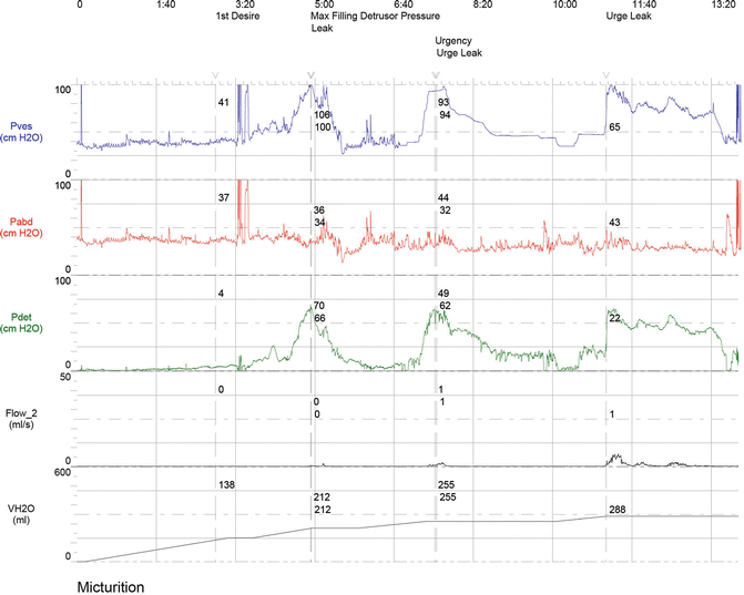
Additionally, the P det during filling should remain low and constant. Rises in P det, not associated with a detrusor contraction, indicate decreased compliance and must be noted. Evaluation of detrusor function is achieved by examining the tracing for the presence of detrusor contractions. Urodynamic detrusor contractions are defined as any significant rise in P det during filling or provocation (Fig. 12.1). A normal or stable tracing should be absent of any detrusor contractions. The presence of detrusor contractions indicates an unstable detrusor that is referred to as detrusor overactivity.


Fig. 12.1
Detrusor overactivity: multichannel cystometry showing detrusor overactivity
As with all urodynamic tests, the results obtained from filling cystometry can be highly variable, depending on various technical aspects (i.e., type of catheters, type of media, filling rate, patient position) and differences in interpretation. Furthermore, the test provides only the results of a single filling cycle. Despite this, it continues to be a frequently utilized test to evaluate patients for stress, urge, or mixed incontinence.
Evaluation of Bladder Emptying or Voiding
Post-void Residual
The simplest evaluation of bladder emptying is to obtain a post-void residual either after passive or active filling of the bladder. Techniques for determining post-void residual (PVR) include ultrasonic techniques (transvaginal, abdominal, and Doppler planimetry) or catheterization. The choice of technique should be based on availability of equipment, diagnostic accuracy, and considerations of patient comfort. While ultrasonic techniques are the least invasive, they represent indirect measures of volume based on measurements on several axis that may introduce error. Direct measurement utilizing a catheter places the patient at some discomfort during the initial placement of catheter, but is generally well tolerated. It has a distinct advantage as it provides the most accurate measure of PVR.
Normal values for post-void residuals are often quoted as values less than 50–100 cc [13, 14]. An isolated finding of an elevated PVR should be interpreted with caution as variation in PVRs can be due to either patient or equipment factors. It is important to ensure that an abnormal PVR is found on repetitive testing. A high PVR often indicates incomplete voiding due to either poor detrusor contractility or outlet obstruction and should prompt further investigation.
Noninvasive Uroflowmetry
Noninvasive uroflowmetry describes a test utilized to calculate the urine flow in relation to voided volume. Patients are asked to void into an electronic volume detector that measures weight changes over time. A graphical representation of the flow pattern is generated for the clinician to interpret. Urine flow is described as continuous or intermittent. Additionally, several objective values are calculated including, flow rate (volume expelled per unit of time), maximum flowrate (Q max), and total voided volume [15].
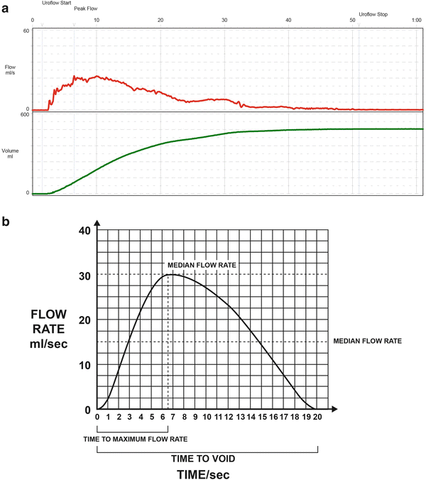
In a normal uroflow, a flow curve generated should rise in amplitude until a high maximum flowrate is achieved and subsequently taper off as micturition is completed. The curve should appear smooth without rapid changes throughout the entire voiding phase (Fig. 12.2).


Fig. 12.2
Uroflow: (a) Actual uroflow showing a normal flow curve. (b) For ease of interpretation, a simplied tracing with variability removed is provided to illustrate how various measurements are obtained
Clinical utility of noninvasive uroflowmetry has been debated, especially in the case of women. The advantage is the relative low cost of the test and potential use as a preliminary screening tool for voiding dysfunction. Interpretation of uroflowmetry is highly dependent on the voided volume. In general, experts agree that in order to properly assess a uroflow study, the patient must void at least 150 cc. Maximum flow rates in women vary with age, sex, and the starting urine volume. Several studies have established maximum urine flow rates to be greater than 12 mL/s [13, 16]. According to the most recent ICS guidelines, abnormally slow flow rates have been determined as under the tenth percentile of the Liverpool nomogram which takes into account the voided volume [17, 18].
Abnormalities in the flow pattern or rate can be due to various pathologies. While specific pathologies that may produce an abnormal uroflow are beyond the scope of this chapter, in general, factors that affect passive relaxation of the bladder neck (i.e., bladder neck obstruction), urethral resistance (i.e., urethral stricture), or detrusor contractility (i.e., neuropathic lesions, pharmacologic manipulation, etc.) will alter the uroflowmetric parameters.
Pressure-Flow Study
Pressure-flow study combines the basic principles of uroflowmetry with the addition of abdominal and bladder pressure measurements. This combination allows evaluation of the pressures that are being generated during the voiding phase, therefore allowing the clinician to examine the pressure volume relationship during micturition. The goal of interpreting a pressure-flow study is to assess whether the neurologic mechanism of micturition is intact. This includes the ability of the detrusor muscle to contract and generate an appropriate voiding pressure, the urethral sphincter to relax, and that the timing of these events are coordinated to provide a smooth continuous flow.
Measurements obtained during a pressure-flow study have been defined by ICS and include premicturition pressure, opening time, opening pressure, maximum pressure, pressure at maximum flow, closing pressure, contraction pressure at maximum flow, and flow delay [3]. While the definitions and interpretations of these measurements are beyond the scope of this chapter, it is important to realize that these additional measurements are possible due to the use of pressure catheters and allow the practitioner to objectively assess for voiding dysfunction.
In general, abnormalities in a pressure-flow study can be categorized into three general groups of pathologies including detrusor contractility issues (i.e., acontractile detrusor), bladder outlet obstructions (i.e., stricture), or neurophysiologic abnormalities (i.e., detrusor sphincter dyssynergia). Evaluation of the voiding P det and flow rate can help the practioner delineate the potential cause. Abnormal pressure-flow studies fall under two general patterns: (1) low flow rate and an elevated P det, suggestive of bladder outlet obstruction; (2) low flow rate and low P det, suggesting potential contractility issue with the detrusor muscle. Several studies have looked to determine specific urodynamic criteria to diagnose bladder outlet obstruction. Furthermore, Blavais and Groutz created a nomogram based on several previous studies and defined bladder outlet obstruction as the presence of a free Q max < 12 mL/s and P det of > 20 cm H2O in a pressure-flow study [19].
While the utility of pressure-flow studies in women have been debated, the most common indications for pressure-flow studies in women include evaluation of obstructed voiding or voiding dysfunction includes preoperative evaluation of patients with pelvic organ prolapse, patients who have undergone previous pelvic surgery, or postoperative patients who have new onset voiding dysfunction. Finally, while it can be performed as an individual test, most often it is combined with filling cystometric studies.
Urethral Function Testing
Urethral Pressure Profile
Urethral pressure profile (UPP) is a test that attempts to measure the urethral pressure throughout the length of the urethra during rest. Variations of the UPP include cough or stress pressure profiles in which the same procedure is performed while the patient coughs. It involves placement of a urethral pressure catheter that will measure the intraluminal pressure as the catheter is withdrawn at a constant rate of speed. While several different catheter types exist, microtransducers are the most commonly used.
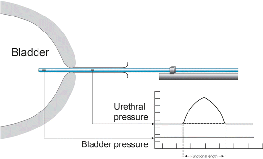
While less commonly used today, the goal of UPP is to distinguish between intrinsic sphincter deficiency (ISD) from stress urinary incontinence (SUI). Several objective measurements including maximum urethral pressure (MUP), urethral closure pressure profile (UCPP), maximum urethral closure pressure (MUCP), functional profile length, and pressure “transmission” ratio are measured in order to assess the urethral closure mechanism. MUCP is calculated by taking the difference between MUP and P ves (Fig. 12.3).


Fig. 12.3
Urethral pressure profile: Illustration showing a normal tracing
The clinical applicability of UPP is limited by lack of standardization of the procedure and in the interpretation of the test itself. Several authors have attempted to correlate lower MUCP with SUI. Other studies have sought to use MUCP to predict surgical success and failure. Several retrospective studies show higher postsurgical failure rates in women with low MUCP, often defined as ≤20 cm H2O [20, 21]. However, a recent review of the role of urodynamic evaluation of SUI showed no clear benefit in improving case selection or surgical technique. The author concluded that urodynamic assessment (LLP and UPP) could not accurately predict the outcome of surgical management of SUI [22].
Leak Point Pressure
The ability of the urethra to prevent the involuntary leakage of urine can be evaluated by measuring the leak point pressure. Under normal physiologic and functional conditions, the urethral resistance should generate enough pressure to compensate for any abdominal or detrusor pressure that would be experienced through normal activities. Leak point pressure is defined as the lowest detrusor (P det) or intravesical pressure (P ves) at which involuntary expulsion of urine from the urethral meatus is observed.
Stay updated, free articles. Join our Telegram channel

Full access? Get Clinical Tree


