117 Trisomy 21
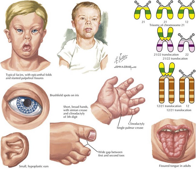
Down syndrome, or trisomy 21, is the most common chromosomal abnormality among live-born infants and is the most frequent microscopically identifiable genetic cause of mental retardation. An extra copy of chromosome 21 results in an overexpression of the genes found on this chromosome to result in the phenotypic differences seen in patients with Down syndrome. This disorder is characterized by a variety of dysmorphic features, congenital malformations, and medical problems (Figures 117-1).
Etiology and Pathogenesis
Although trisomy 21 is responsible for 1 : 150 first-trimester spontaneous abortions, the live-born incidence is approximately 1 : 800-1 : 1000, and its incidence is highly correlated with advanced maternal age. The additional copy of chromosome 21 usually occurs from meiosis I nondisjunction, when the pair of homologous chromosomes 21 fail to separate. Errors that occur during meiosis are nearly always maternal in origin, and the incidence increases with advancing maternal age. This is likely attributable to the years from oocyte formation during maternal embryogenesis to the time of fertilization that the oocyte spends suspended in meiosis I.
Although trisomy of chromosome 21 (e.g., 47,XY, +21) in which a complete extra copy of chromosome 21 is present, causes approximately 94% of cases, other causes are also seen. Approximately 2% to 3% of cases of trisomy 21 can present with mosaicism (e.g., 47,XY, +21 [50%] or 46,XY[50%]) in which there is a mixture of trisomic cells and normal cells. This can lead to an attenuated phenotype, but it depends on the relative mosaicism of different tissues. Finally, approximately 3% to 4% of cases are caused by a Robertsonian translocation, in which the q arm of chromosome 21 is translocated onto another chromosome. This results in a fetus with 46 chromosomes but three copies of the long arm of chromosome 21, which carries all of the functional genes of this chromosome (e.g., 46,XY,der(15 : 21)(q10;q10), +21; Figure 117-1). Although most Robertsonian translocations are new mutations and occur regardless of maternal age, it is important to distinguish the cause and rule out a translocation carrier parent, which substantially increases the risk of recurrence.
Clinical Presentation and Differential Diagnois
The diagnosis of trisomy 21 is often made by prenatal screening. When no prenatal diagnosis has been made, Down syndrome is usually recognized from the characteristic phenotype present in the newborn. The diagnosis is confirmed with a karyotype.
Phenotypic Features
The pathogenesis of the characteristic appearance of infants with trisomy 21 and associated malformations of Down syndrome are presumably related to dosage effects of genes on chromosome 21, specifically those in a 5Mb region on 21q22. Certain malformations occur more frequently than others, thus supporting the concept that certain genes rather than a specific embryologic event are involved. The diagnosis of Down syndrome is often straightforward and is based on the overall appearance and characteristics of individuals. Neonates with Down syndrome are typically hypotonic and have poor developmental reflexes. Neonates can also have redundant nuchal skin folds. The skull may be mildly microcephalic with a small occiput and large fontanelles. The eyes are almond shaped with epicanthal folds and upslanting palpebral fissures, and the irises may demonstrate Brushfield spots. The nose is usually short with a low nasal bridge. Usually, the mouth is downturned, and because the oral cavity is small, the tongue often protrudes. The ears may be low set with an overfolded superior helix. The hands are short and commonly have the characteristic single palmar crease.
Developmental Impairment
Nearly all individuals with Down syndrome have some degree of cognitive impairment, although the range is quite wide. Most affected are mildly to moderately mentally retarded, with IQ in the 50 to 70 or 35 to 50 ranges, respectively. Some may be severely impaired with IQ ranges from 20 to 35. Cognitive impairment is evident within the first year of life. Developmental milestones are met typically at twice the age compared with the average population with sitting at around 11 months, creeping at around 17 months, and walking around 26 months. The sequence of the development of language is the same, but the rate is much slower. First words are spoken on average at around 18 months. Affected children will still gain new skills, but the IQ tends to decline through childhood and plateaus in adolescence.
In more than 60% of children, the profile of cognitive impairment is one in which language comprehension is equal to mental age, and language production is more delayed. However, in about a third of children with Down syndrome, language comprehension, mental age, and language production are equal.
Comorbidities
Several important comorbidities are associated with Down syndrome. Among these, it is important to recognize heart disease, gastrointestinal abnormalities, growth problems, eye problems, hearing loss, hematologic disorders, immune deficiency, endocrine disorders, reproductive disorders, atlantoaxial instability (AAI), sleep apnea, skin disorders, and behavior disorders.
Heart Disease
Approximately 50% of children with Down syndrome have congenital heart disease that includes atrioventricular septal (ASDs), atrioventricular canal or endocardial cushion defects (45%), ventricular septal defects (35%), isolated secundum ASDs, (8%), isolated patent ductus arteriosus (7%), and tetralogy of Fallot (4%). Furthermore, asymptomatic children without congenital structural heart disease may develop valve abnormalities, including mitral valve prolapse, mitral regurgitation, and aortic regurgitation.
Gastrointestinal Disease
Gastrointestinal tract abnormalities occur in about 5% of children with trisomy 21 with the most common being duodenal atresia or stenosis (2.5%), sometimes occurring with an annular pancreas. The characteristic radiographic finding associated with duodenal atresia or stenosis is the “double-bubble” sign. Other anomalies, although less frequently seen, include imperforate anus and esophageal atresia with tracheoesophageal fistula. Conversely, nearly 30% of patients with duodenal atresia or stenosis and 20% with annular pancreas have Down syndrome. Hirschsprung disease is more common in trisomy 21 compared with the general population, although the risk is less than 1%. Trisomy 21 appears to be strongly associated with celiac disease with an incidence of between 5% and 16%, which is five- to 16-fold greater than the general population.
Growth and Stature
Head circumference, length, and weight are all lower in infants and children with trisomy 21 than unaffected children. Compared with their siblings, at birth, children with trisomy 21 weigh on average 0.2 to 0.4 kg less. The rate of growth of children with trisomy 21 is reduced, especially during infancy and adolescence, and is more severely reduced in children with congenital heart disease. In adolescence, the growth spurt of affected children occurs earlier, further blunting the ultimate height. In adults with Down’s syndrome, the average height in men and women is 61.7 and 57 inches (157 and 144 cm), respectively, and the average weight is 157 and 140 lb (71 and 64 kg), respectively.
The cause of trisomy 21–associated growth retardation remains unknown. In some patients, low serum levels of insulin-like growth factor 1 (IGF-1) and lower spontaneous and induced secretion of growth hormone (GH) have been reported. Hypothalamic dysfunction may lead to suboptimal endogenous GH production. Selective deficiency of IGF-1 has been seen in individuals with Down syndrome who are older than the age of 2 years.

Stay updated, free articles. Join our Telegram channel

Full access? Get Clinical Tree

