Thyroid Disease

68 Thyroid Disease


The follicular cells of the thyroid gland produce triiodothyronine (T3) and thyroxine (T4), thyroid hormones that have important roles in growth and development, as well as influential effects on metabolism, the central nervous system (CNS), and the cardiovascular system. This chapter reviews thyroid physiology, as well as the most common childhood thyroid disorders.


Thyroid Hormone Production


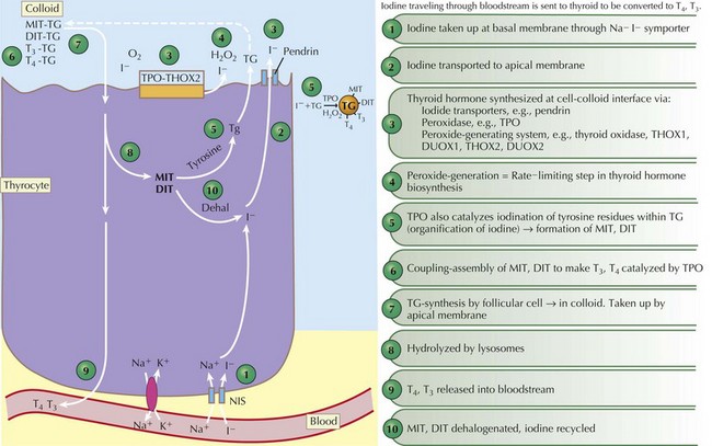
Thyroid hormones are produced by the coupling of iodine molecules to the amino acid tyrosine (Figure 68-1). The principal secreted thyroid hormone is T4. The normal thyroid secretes only small amounts of T3, and most circulating T3 (≈70%-90%) is derived from peripheral deiodination of T4. Thyroid hormones circulate bound to carrier proteins, including thyroid-binding globulin (TBG), transthyretin (thyroxine-binding prealbumin), and albumin. Less than 1% of circulating T4 and T3 are unbound or “free.”


Hypothalamic thyrotropin-releasing hormone (TRH) stimulates thyrotrope cells in the anterior pituitary to release thyroid-stimulating hormone (TSH), which stimulates production and release of thyroid hormone. When T4 levels are inadequate, a negative feedback loop activates the hypothalamic–pituitary axis and results in increased secretion of TSH, which then acts on the thyroid gland to stimulate increased hormone synthesis. When circulating levels of T4 and T3 are high, as in Graves’ disease or overtreatment with exogenous T4, this negative feedback loop acts to reduce or suppress TSH secretion. Circulating levels of T4 are also influenced by peripheral conversion to either T3 or reverse T3 (rT3), an inactive form of thyroid hormone.


Circulating T4 and T3 enter cells by diffusion and carrier-mediated transport processes; inside the cell, T4 is then converted to T3. T3 is the most active thyroid hormone because it binds thyroid hormone receptors with approximately 10 times the affinity of T4. The T3–receptor complex is transported to the nucleus and regulates transcription of a variety of genes, ultimately leading to the synthesis of proteins that manifest thyroid hormone action in peripheral tissues. T4 is necessary for normal growth and development and is absolutely critical for brain development in utero as well as during the first 2 years of life.


Congenital Hypothyroidism


Etiology and Pathogenesis


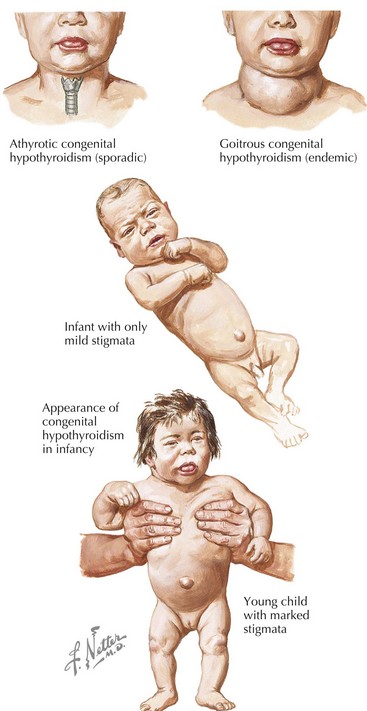
Congenital hypothyroidism (CH) is one of the most common causes of preventable mental retardation (Figure 68-2). Fortunately, early identification and rapid treatment lead to normal neurocognitive development. Although approximately 10% of cases of CH are transient and caused by factors such as iodine exposure, prematurity, or maternal transfer of antithyroid antibodies, in most cases, hypothyroidism is permanent. Worldwide, iodine deficiency is the most common cause of CH. However, in areas of the world where iodine deficiency is uncommon, CH most commonly results from thyroid dysgenesis (≈75% of cases); thyroid dyshormonogenesis (≈10%), TSH deficiency (5%), and genetic defects in the TSH receptor are much less common. The incidence of CH is approximately one in 3000 to 4000 births. In most cases, CH is sporadic, but mutations in genes encoding transcription factors that are required for normal development of the thyroid gland are present in about 10% to 15% of cases. Circulating levels of TSH are elevated and thyroid hormone levels are low in those with CH.


A clinical picture that is similar to CH can occur in children who have genetic defects in the thyroid hormone receptor or in the MCT8 protein required to transport thyroid hormone into cells. Unlike CH, however, these patients have elevated serum levels of both TSH and thyroid hormones.


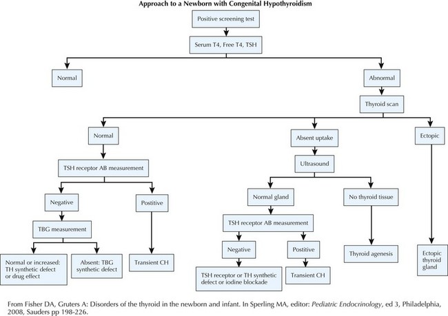
Evaluation (Figure 68-3)


NBS for CH is based on collection of a blood sample from the newborn between 48 hours and 4 days of life. Earlier measurement may lead to erroneous results because of the normal physiologic surge in TSH that occurs soon after birth. NBS programs use blood spots collected on filter paper and use one of two strategies to identify infants with CH: a primary TSH with backup T4 or a primary T4 with a backup TSH method. An abnormal result should be further evaluated immediately using serum-based assays for TSH and T4 or free T4. Premature or ill infants may have false-negative or false-positive results and should be retested by 7 days of age. Any infant with a TSH level above 40 mU/L with low T4 is considered to have primary hypothyroidism. In addition to T4 and TSH measurements, a thyroglobulin (TG) level may be helpful because elevation suggests dyshormonogenesis.


When there is history of maternal autoimmune thyroid disease, measurement of TSH–receptor binding antibodies in the infant or the mother may identify transient CH. Other diagnostic tests include thyroid ultrasonography and technetium or iodine (I-123) scans to identify functional thyroid tissue, as well as the perchlorate washout test to detect iodine organification defects that might indicate Pendred’s syndrome. Treatment with levothyroxine (L-T4) should not be delayed to perform imaging.


Treatment


To avoid neurocognitive deficit, newborns with CH must be treated promptly with L-T4 and monitored closely by a pediatric endocrinologist. L-T4 is instituted at a dosage of 10 to 17 µg/kg/d initially, with a goal to normalize the serum T4 level within 2 weeks (fT4 >2 ng/dL) and serum TSH by 1 month of age. There are no suitable liquid preparations of L-T4, so tablets must be used. Tablets should be crushed and mixed with a few milliliters of formula, breast milk, or water. Soy-based formula, fiber, or iron may reduce absorption of L-T4 and should be given separately.


Serum levels of T4, T4 index or free T4, and TSH should be measured every 1 to 2 months during the first 6 months of life, every 3 to 4 months until 3 years of age, and then every 6 to 12 months until growth is complete. The half-life of T4 in the circulation is 1 week, so levels of TSH and T4 should be repeated 4 to 5 weeks after dose changes to ensure appropriate steady-state levels. An appropriately treated child will have serum T4 levels that are at or above the upper limit of normal with a serum TSH level of 1 to 2 mU/mL. The serum TSH level is not a reliable indicator of euthyroidism in children with pituitary or hypothalamic disorders, and in these patients, the T4 level should be maintained at the upper limit of the assay’s normal range. Infants with suspected transient hypothyroidism should continue L-T4 therapy until at least age 2 years, when thyroid-dependent CNS myelinization is complete.

< div class='tao-gold-member'>

Stay updated, free articles. Join our Telegram channel

Jun 19, 2016 | Posted by in PEDIATRICS | Comments Off on Thyroid Disease

Full access? Get Clinical Tree

Get Clinical Tree app for offline access