CHAPTER 50 The mechanism of continence
Urinary Incontinence
Introduction
Urinary incontinence may be defined as a condition in which an involuntary loss of urine is a social or hygienic problem and is objectively demonstrable. Therefore, continence is the ability to retain urine within the bladder between voluntary acts of micturition. In order to fully comprehend the pathological processes which lead to the development of urinary incontinence, a clear understanding of normal mechanisms for the maintenance of continence is fundamental.
Anatomy of the lower urinary tract
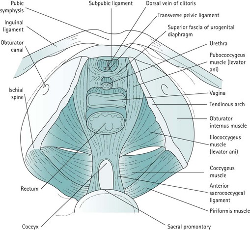
The bladder
The bladder consists of three layers of smooth muscle, known collectively as the ‘detrusor’, which functions as one syncytial mass with the exception of the trigone. The outer smooth muscle layer is oriented longitudinally, the middle layer is oriented circularly and the inner layer is oriented longitudinally.
Histochemically, the detrusor muscle has been shown to be rich in acetylcholinesterase by virtue of its rich cholinergic parasympathetic nerve supply. In contrast, the trigone only comprises two layers of smooth muscle. The inner layer is similar to the rest of the detrusor, but the outer layer consists of smooth muscle bundles with a sparse parasympathetic nerve supply. The outer layer extends to, and is continuous with, the proximal urethra and the distal portion of the ureter, where it may have a role in preventing ureteric reflux. The smooth muscle that forms the bladder neck is separate from the detrusor, with little or no sphincteric effect. Two or three layers of transitional epithelia cover the detrusor and secrete a protein on to their luminal surface that forms a watertight blood–bladder barrier. When empty, the urothelium relaxes into numerous folds or rugae.
The urethra
The normal female urethra is 30–50 mm in length. It comprises smooth and striated muscle. The smooth muscle is continuous with that of the detrusor, but has minimal parasympathetic innervation and little sphincteric effect in contrast to the profusion of acetylcholinesterase within the detrusor cells. The external urethral sphincter (rhabdosphincter) is one of two striated muscle components surrounding the urethra. Its fibres are bulked anteriorly at the midurethral level, are slow twitch in nature and are involved in maintaining continence at rest. The second striated muscle component is the periurethral portion of the levator ani, which is separated from the external urethral sphincter by a connective tissue septum. The fibres are bulked anterolaterally at a lower level than the external urethral sphincter, are fast twitch in nature and are involved in maintaining continence under stress.
The mucosa of the urethra is lined by pseudostratified transitional epithelia proximally, changing to non-keratinized stratified squamous epithelia distally. The junction between the two cell types alters with age and oestrogenic status, which may affect urinary symptoms. In young females, the submucosa has a rich venous supply which engorges the tissues, helping to close the urethra. This ceases after the menopause, and may be involved in the development of stress incontinence later in life due to poor urethral closure.
The pelvic floor
For the context of this chapter, the pelvic floor structures that will be described are the levator ani muscles, the endopelvic fascia and condensations of this fascia which form the ligaments. The levator ani muscles are often described as funnel-shaped structures, but in-vivo imaging has revealed that they are, in fact, more horizontal. The fibres of each side pass downwards and inwards. The two muscles, one on each side, constitute the pelvic diaphragm. Defects in the levator ani allow the urethra, vagina and rectum to pass. The levator ani is subdivided into the pubococcygeus, iliococcygeus and coccygeus. The pubococcygeus arises anteriorly from the posterior aspect of the pubic bone and from the anterior portion of the arcus tendineus (white line), which is a condensation of the obturator internus fascia. The medial fibres of the pubococcygeus merge with the fibres of the vagina and perineal body, and have been given various names such as puborectalis, pubourethralis and pubovaginalis. The iliococcygeus arises from the remainder of the arcus tendineus, partly overlapping the pubococcygeus on its perineal surface and extending to the medial surface of the ischial spine. The coccygeus or ischiococcygeus is a rudimentary muscle arising from the tip of the ischial spine, and quite often constitutes only a few muscle fibres on the sacrospinous ligament. Posteriorly, the muscles of the levator ani or pelvic diaphragm insert into the sides of the coccyx and the anococcygeal raphe, which is formed by the interdigitation of muscle fibres from either side. The most medial fibres of the pubococcygeus that pass round the rectum at the anorectal junction form the puborectalis muscle (Figure 50.1).
The endopelvic fascia invests all the structures that lie within the pelvis and is mainly composed of loose areolar tissue, although smooth muscle cells have been identified. The layer between the bladder, urethra and vagina is termed the ‘vesicovaginal fascia’, and the layer between the vagina and rectum is known as the ‘rectovaginal fascia’ or ‘rectovaginal septum’. Laterally, both these layers attach to a condensation of fascia known as the ‘arcus tendineus fasciae pelvis’. This is a condensation of the endopelvic fascia which runs from the posterior part of the pubic ramus to the ischial spine, and attaches to the stronger underlying obturator internus muscle fascia. The urethra has an intimate attachment to the lower one-third of the vagina, and therefore the supports for these structures are identical. The urethra has a further condensation of the endopelvic fascia which attaches to the symphysis pubis. These are known as the ‘pubourethral ligaments’ and lie at the midportion of the urethra. They are fairly loose attachments, and allow movement of the bladder neck during straining and also by voluntary contraction of the levator ani muscles.
The uterus, cervix and upper third of the vagina are attached to the pelvic side wall by broad condensations of endopelvic fascia with a high content of smooth muscle cells, known as the ‘cardinal and uterosacral ligaments’. These ligaments originate from the region of the greater sciatic foramen and the lateral aspects of the sacrum. In the erect position, these ligaments run almost vertically and suspend their attached structures.
The levator ani muscles, ligaments and endopelvic fascia work in synergy. If any of these structures is not intact, the mechanisms of support and continence may be affected.
Innervation of the bladder and urethra
The bladder has a rich parasympathetic nerve supply (Figure 50.2). The postganglionic cell bodies lie either in the bladder wall or pelvic plexuses, innervated by preganglionic fibres that originate from cell bodies in the grey columns of S2–4. There is little sympathetic innervation of the bladder, although greater quantities of noradrenergic terminals can be detected in the bladder neck or trigone. Noradrenergic effects can be either inhibitory or excitatory, depending on the receptor type present.

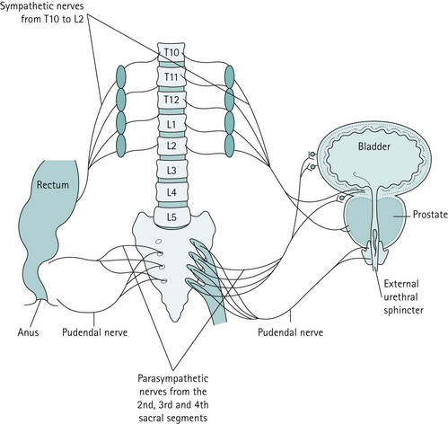
Figure 50.2 The major innervation pathways at the pelvic level that are basic to control of micturition and continence.
The cell bodies of the sympathetic nerves originate in the grey matter of T10–L2, and pass through the sympathetic chain via the lumbar splanchnic nerve and left and right hypogastric nerves to the pelvic plexus. It is thought that the sympathetic nervous system exerts its effects by inhibiting the parasympathetic nervous system rather than by direct action. The visceral nerve afferents pass along the sacral and thoracolumbar visceral efferents, relaying sensations of touch, pain and distension. However, transection has little or no effect on micturition. The urethra possesses similar autonomic innervation. Parasympathetic efferents cause contraction but the functional significance of this remains in doubt. There is no obvious sphincteric function, but contraction produces shortening and widening of the urethra along with detrusor contraction during micturition. Sympathetic efferents innervate the predominantly α-adrenoreceptors.
Innervation of striated muscle
The rhabdosphincter muscle is supplied via the pelvic splanchnic nerves travelling with the parasympathetic fibres to the intrinsic smooth muscle of the urethra. This is analogous to the puborectalis muscle.
The extrinsic periurethral muscle of the levator ani is innervated by motor fibres of the perineal branch of the pudendal nerve. This is in common with the external anal sphincter (EAS). The pudendal nerve also supplies the striated elements of the levator ani on both sides, and there are associated somatic afferent fibres which travel with the pudendal nerves. They ascend via the dorsal columns to convey proprioception from the pelvic floor.
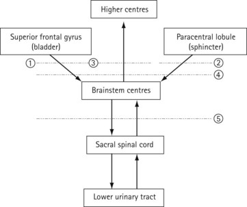
Central nervous control of continence
The connections of the lower urinary tract within the central nervous system are complex (Figure 50.3). There are many discrete areas which influence micturition, and these have been identified within the cerebral cortex, in the superior frontal and anterior cingulate gyri of the frontal lobe and the paracentral lobule; within the cerebellum, in the anterior vermis and fastigial nucleus; and in subcortical areas, including the thalamus, the basal ganglia, the limbic system, the hypothalamus and discrete areas of the mesencephalic pontine medullary reticular formation. The full function and interactions of these various areas are incompletely understood, although the effects of ablation and tumour growth in humans and stimulation studies in animals have given some insights.

Figure 50.3 The interaction of the nervous system in micturition. Locations of possible nervous lesions are denoted by numbers. 1, lesions isolating superior frontal gyrus will prevent voluntary postponement of voiding; 2, lesions isolating paracentral lobule, often with hemiparesis, will cause spasticity of urethral sphincter and retention; 3, pathways of sensation are not known accurately; in theory, isolated lesion of sensation above brainstem would lead to unconscious incontinence, and defective conduction of sensory information would explain enuresis; 4, lesions above brainstem centres lead to involuntary voiding that is coordinated with sphincter relaxation; 5, lesions below brainstem centres but above the sacral spinal cord lead, after a period of bladder paralysis, to involuntary ‘reflex’ voiding that is not coordinated with sphincter relaxation.
The centres within the cerebral cortex are important in the perception of sensation in the lower urinary tract, and the inhibition and subsequent initiation of voiding. Lesions in the superior frontal gyrus and the adjacent anterior cingulate gyrus reduce or abolish both the conscious and unconscious inhibition of the micturition reflex. The bladder tends to empty at low functional capacity. Sometimes, the patient is aware of the sensation of urgency; sometimes, micturition may be entirely unconscious. These areas, which have been localized by functional magnetic resonance imaging (MRI), are supplied by the anterior cerebral and pericallosal arteries; spasm or occlusion of these arteries produces incontinence.
Localized lesions more posteriorly in the paracentral lobule may produce retention rather than incontinence because of the combination of impaired sensation and spasticity of the pelvic floor.
The thalamus is the principal relay centre for pathways projecting to the cerebral cortex, and ascending pathways activated by bladder and urethral receptors synapse on neurones in specific thalamic nuclei which have reciprocal connections with the cortex. Electrical stimulation of the basal ganglia in animals leads to suppression of the detrusor reflex, whereas ablation has resulted in detrusor hyper-reflexia; patients with Parkinsonism are commonly shown to have detrusor instability on cystometric examination.
Within the pontine reticular formation are two closely related areas with inhibitory and excitatory effects on the sacral micturition centre in the conus medullaris. Lesions of the cord below this level always lead to incoordinate voiding with a failure of urethral relaxation during detrusor contraction; lesions above this level may be associated with normal although involuntary micturition.
The mechanism of urinary continence
The mechanism of urinary continence is a complex dynamic process which relies on intact fascia, ligaments and muscles with their accompanying nerve and vascular supply. It relies on maximum urethral pressure being higher than maximum detrusor pressure. There are special features of physiology and anatomy that contribute to the maintenance of a low detrusor pressure, and adequate urethral closure and positioning. These features are discussed below.
Bladder
An intravesical pressure that exceeds urethral pressure will lead to incontinence. There are a number of factors which maintain a low intravesical pressure.
The hydrostatic pressure at the bladder neck
Any fluid within the bladder will itself have a pressure. This is due to a vertical gravitational pressure gradient. For clinical purposes, the intravesical pressure defines the pressure in the bladder with respect to atmospheric pressure measured at the level of the upper border of the symphysis pubis. This pressure works against the mechanisms that produce urethral closure, and very rarely amounts to more than 10 cmH2O.
Transmission of intra-abdominal pressure
The bladder is normally an intra-abdominal organ and is subject to the pressures of adjacent organs and abdominal pressure. The pressures are normally equally transmitted to the bladder, the bladder neck and proximal areas of the urethra, and this helps to maintain continence.
Tension in the bladder wall
Due to higher control of the basic visceral reflexes, the bladder does not contract in response to stretch receptors within the bladder wall. This and the actual ability of the bladder to be compliant (i.e. to allow for a large rise in volume and little rise in pressure) make it an ideal storage organ.
Urethra
There are several components of urethral function that are necessary to maintain good urethral closure.
The hermetic seal
The vascularity in the submucosal urethral plexuses has been mentioned above, and there are also secretions that allow this seal to be maintained. This may fail after the menopause.
The intrinsic smooth and striated muscles and the extrinsic striated muscles
The intrinsic smooth and striated muscles exert a constant pressure from their constant tone, and the extrinsic striated muscle contracts during moments of stress to maintain urethral closure. It has been estimated from urethral pressure studies that resting urethral pressure is one-third due to external striated muscle effects, one-third due to smooth muscle effects and one-third due to its vascular supply. The highest pressure is found at the midurethral point.

Full access? Get Clinical Tree

