Chapter 666 The Foot and Toes
The foot may be divided into the forefoot (toes and metatarsals), the midfoot (cuneiforms, navicular, cuboid), and the hindfoot (talus and calcaneus). Although the tibiotalar joint (ankle) provides plantar flexion and dorsiflexion, the subtalar joint (between the talus and calcaneus) is oriented obliquely, providing inversion and eversion. Inversion represents a combination of plantar flexion and varus, and eversion involves dorsiflexion and valgus. The subtalar joint is especially important for walking on uneven surfaces. The talonavicular and calcaneocuboid joints connect the midfoot with the hindfoot.
Abnormalities affecting the osseous and articular structures of the foot may be congenital, developmental, neuromuscular, or inflammatory. Problems with the foot and/or toes may be associated with a host of connective tissue diseases and syndromes such as overuse syndromes, which are commonly observed in young athletes. Symptoms can include pain and abnormal shoe wear; cosmetic concerns are common.
666.1 Metatarsus Adductus
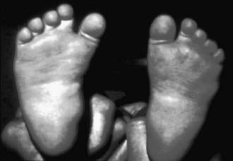
Metatarsus adductus is common in newborns and involves adduction of the forefoot relative to the hindfoot. When the forefoot is supinated and adducted, the deformity is termed metatarsus varus (Fig. 666-1). The most common cause is intrauterine molding, where the deformity is bilateral in 50% of cases. As with other intrauterine positional foot deformities, a careful hip examination should always be performed.
Clinical Manifestations
The forefoot is adducted (occasionally supinated), whereas the midfoot and hindfoot are normal. The lateral border of the foot is convex, and the base of the 5th metatarsal appears prominent. Range of motion at the ankle and subtalar joints is normal. Both the magnitude and the degree of flexibility should be documented. When the foot is viewed from the plantar surface, a line through the midpoint of (and parallel to) the heel should normally extend through the 2nd toe. Flexibility is assessed by stabilizing the hindfoot and midfoot in a neutral position with one hand and applying pressure over the 1st metatarsal head with the other. In the walking child with an uncorrected metatarsus adductus deformity, an in-toe gait and abnormal shoe wear can occur. A subset of patients also have a dynamic adduction deformity of the great toe (hallux varus), which is often most noticeable during ambulation. This usually improves spontaneously and does not require treatment.
Radiographic Evaluation
Radiographs are not performed routinely, but an anteroposterior (AP) and lateral weight-bearing or simulated weight-bearing radiographs are indicated in toddlers or older children with residual deformities. The AP radiographs demonstrate adduction of the metatarsals at the tarsometatarsal articulation and an increased intermetatarsal angle between the 1st and 2nd metatarsals.
Treatment
The treatment of metatarsus adductus is based on the rigidity of the deformity; most children respond to nonoperative treatment. Deformities that are flexible and overcorrect into abduction with passive manipulation may be observed. The feet that correct just to a neutral position can benefit from stretching exercises and retention in a slightly overcorrected position by a splint or reverse-last shoes. These are worn full time (22 hr/day), and the condition is re-evaluated in 4-6 wk. If improvement occurs, treatment can be continued. If there is no improvement, serial plaster casts should be considered. When stretching a foot with metatarsus adductus, care should be taken to maintain the hindfoot in neutral to slight varus alignment to avoid creating hindfoot valgus. Feet that cannot be corrected to a neutral position might benefit from serial casting; the best results are obtained when treatment is started before 8 mo of age. In addition to stretching the soft tissues, the goal is to alter physeal growth and stimulate remodeling, resulting in permanent correction. Once flexibility and alignment are restored, orthoses or corrective shoes are generally recommended for an additional period. A dynamic hallux varus usually improves spontaneously, and no active treatment is required.
Surgical treatment may be considered in the small subset of patients with symptomatic residual deformities that have not responded to previous treatment. Surgery is generally delayed until children are 4-6 yr of age. Cosmesis is often a concern, and pain and/or the inability to wear certain types of shoes occasionally leads patients to consider surgery. Options for surgical treatment include either soft-tissue release or osteotomy. An osteotomy (midfoot or multiple metatarsals) is most likely to result in permanent restoration of alignment.
666.2 Calcaneovalgus Feet
Harish S. Hosalkar, David A. Spiegel, and Richard S. Davidson
The calcaneovalgus foot is common in the newborn (30-50% of newborns have a mild version) and is secondary to in utero positioning. Excessive dorsiflexion and eversion are observed in the hindfoot, and the forefoot may be abducted. An associated external tibial torsion may be present.
Clinical Manifestations
The infant typically presents with the foot dorsiflexed and everted, and occasionally the dorsum is in contact with the anterolateral surface of the lower leg. Plantar flexion and inversion are often restricted. As with other intrauterine positional deformities, a careful hip examination should be performed; if there is any concern, hip ultrasonography should be considered. The calcaneovalgus foot may be confused with a congenital vertical talus and is rarely associated with a posteromedial bow of the tibia. A calcaneovalgus deformity may also be seen in older patients, typically those with a neuromuscular imbalance involving weakness or paralysis of the gastrocnemius-soleus complex (polio, myelomeningocele).
Radiographic Evaluation
Radiographs are usually not required, but they should be ordered if the deformity fails to correct spontaneously or with early treatment. AP and lateral radiographs, with a lateral radiograph of the foot in maximal plantar flexion, might help distinguish calcaneovalgus from a vertical talus. If a posteromedial bow of the tibia is suspected, anteroposterior and lateral radiographs of the tibia and fibula are necessary.
Treatment
Mild cases of calcaneovalgus foot, in which full passive range of motion is present at birth, require no active treatment. These usually resolve within the 1st weeks of life. A gentle stretching program, focusing on plantar flexion and inversion, is recommended for cases with some restriction in motion. For cases with a greater restriction in mobility, serial casts may be considered to restore motion and alignment. Casting is rarely required in the treatment of calcaneovalgus feet. The management for cases associated with a posteromedial bow of the tibia is similar.
666.3 Talipes Equinovarus (Clubfoot)
Talipes equinovarus (also known as clubfoot) describes a deformity involving malalignment of the calcaneotalar-navicular complex. Components of this deformity may be best understood using the mnemonic CAVE (cavus, adductus, varus, equinus). Although this is predominantly a hindfoot deformity, there are plantar flexion (cavus) of the 1st ray and adduction of the forefoot/midfoot on the hindfoot. The hindfoot is in varus and equinus. The clubfoot deformity may be positional, congenital, or associated with a variety of underlying diagnoses (neuromuscular or syndromic).
The positional clubfoot is a normal foot that has been held in a deformed position in utero and is found to be flexible on examination in the newborn nursery. The congenital clubfoot involves a spectrum of severity, while clubfoot associated with neuromuscular diagnoses or syndromes are typically rigid and more difficult to treat. Clubfoot is extremely common in patients with myelodysplasia and arthrogryposis (Chapter 674).
Clinical Manifestations
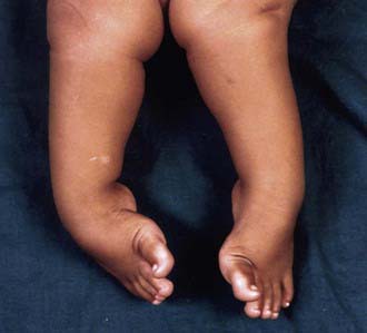
A complete physical examination should be performed to rule out coexisting musculoskeletal and neuromuscular problems. The spine should be inspected for signs of occult dysraphism. Examination of the infant clubfoot demonstrates forefoot cavus and adductus and hindfoot varus and equinus (Fig. 666-2). The degree of flexibility varies, and all patients exhibit calf atrophy. Both internal tibial torsion and leg-length discrepancy (shortening of the ipsilateral extremity) are observed in a subset of cases.
Radiographic Evaluation
Anteroposterior and lateral radiographs are recommended, often with the foot held in the maximally corrected position. Multiple radiographic measurements can be made to describe malalignment between the tarsal bones. The navicular bone does not ossify until 3-6 yr of age, so the focus of radiographic interpretation is the relationships between segments of the foot. A common radiographic finding is “parallelism” between lines drawn through the axis of the talus and the calcaneus on the lateral radiograph, indicating hindfoot varus. Many clinicians believe that radiographs are not required in the evaluation and treatment of clubfoot in infancy and reserve these studies for older children with persistent or recurrent deformities.
Treatment
Nonoperative treatment is initiated in all infants and should be started as soon as possible following birth. Techniques have included taping and strapping, manipulation and serial casting, and functional treatment. Historically, a significant percentage of patients treated by manipulation and casting required a surgical release, which was usually performed between 3 and 12 mo of age. Although many feet remain well aligned after surgical releases, a significant percentage of patients have required additional surgery for recurrent or residual deformities. Stiffness remains a concern at long-term follow-up. Pain is uncommon in childhood and adolescence, but symptoms can appear during adulthood. These concerns have led to considerable interest in less-invasive methods for treating the deformity.
The Ponseti method of clubfoot treatment involves a specific technique for manipulation and serial casting and may be best described as minimally invasive rather than nonoperative. The order of correction follows the mnemonic CAVE. Weekly cast changes are performed, and 5 to 10 casts are typically required. The most difficult deformity to correct is the hindfoot equinus, for which ~90% of patients require an outpatient percutaneous tenotomy of the heel cord. Following the tenotomy, a long leg cast with the foot in maximal abduction (70 degrees) and dorsiflexion is worn for 3 wk; the patient then begins a bracing program. An abduction brace is worn full time for 3 mo and then at nighttime for 3-5 yr. A subset of patients require transfer of the tibialis anterior tendon to the middle cuneiform for recurrence. Although most patients require some form of surgery, the procedures are minimal in comparison with a surgical release, which requires capsulotomy of the major joints (and lengthening of the muscles) to reposition the joints in space. The results of the Ponseti method are excellent at up to 40 yr of follow-up. Compliance with the splinting program is essential; recurrence is common if the brace is not worn as recommended. Functional treatment, or the French method, involves daily manipulations (supervised by a physical therapist) and splinting with elastic tape, as well as continuous passive motion (machine required) while the baby sleeps. Although the early results are promising, the method is labor intensive, and it remains unclear whether the technique will achieve greater popularity in the USA. These minimally invasive methods are most successful when treatment is begun at birth or during the first few months of life.
Surgical realignment has a definite role in the management of clubfeet, especially in the minority of congenital clubfeet that have failed nonoperative or minimally invasive methods, and for the neuromuscular and syndromic clubfeet that are characteristically rigid. In such cases, nonoperative methods such as the Ponseti technique are potentially of value in decreasing the magnitude of surgery required. Common surgical approaches include a release of the involved joints (realign the tarsal bones), a lengthening of the shortened posteromedial musculotendinous units, and usually pinning of the foot in the corrected position. The specific procedure is tailored to the unique characteristics of each deformity. For older children with untreated clubfeet or those in whom a recurrence or residual deformity is observed, bony procedures (osteotomies) may be required in addition to soft-tissue surgery. Triple arthrodesis is reserved as salvage for painful, deformed feet in adolescents and adults.
Alvarado DM, Aferol H, McCall K, et al. Familial isolated clubfoot is associated with recurrent chromosome 17q23.1q23.2 microduplications containing TBX4. Am J Hum Genetics. 2010;87:154-160.
Bridgens J, Kiely N. Current management of clubfoot (congenital talipes equinovarus). BMJ. 2010;340:308-312.
Canto MJ, Cano S, Palau J, et al. Prenatal diagnosis of clubfoot in low-risk populations: associated anomalies and long-term outcome. Prenatal Diag. 2008;28:343-346.
Chang CH, Kumar SJ, Riddle EC, et al. Macrodactyly of the foot. J Bone Joint Surg Am. 2002;84:1189-1194.
Dobbs MB, Gurnett CA. Update on clubfoot: etiology and treatment. Clin Orthop Relat Res. 2009;467:1146-1153.
Herzenberg JE, Radler C, Bor N. Ponseti versus traditional methods of casting for idiopathic clubfoot. J Pediatr Orthop. 2002;22:517-521.
Ippolito E, Fraracci L, Farsetti P, et al. The influence of treatment on the pathology of clubfoot. CT study at maturity. J Bone Joint Surg Br. 2004;86:574-580.
Morcuende JA, Dolan LR, Dietz FR, et al. Radical reduction in the rate of extensive corrective surgery for clubfeet using the Ponseti method. Pediatrics. 2004;113:376-380.
Noonan KJ, Richards BS. Nonsurgical management of idiopathic clubfoot. J Am Acad Orthop Surg. 2003;11:392-402.
Offerdal K, Jebens N, Blaas HGK, et al. Prenatal ultrasound detection of talipes equinovarus in a non-selected population of 49 314 deliveries in Norway. Ultrasound Obstet Gynecol. 2007;30:838-844.
Paton RW, Choudry Q. Neonatal foot deformities and their relationship to developmental dysplasia of the hip. J Bone Joint Surg Br. 2009;91:B655-B658.
Roye DPJr, Roye BD. Idiopathic congenital talipes equinovarus. J Am Acad Orthop Surg. 2002;10:239-248.
Sankar WN, Weiss J, Skaggs DL. Orthopaedic conditions in the newborn. J Am Acad Orthop Surg. 2009;17:112-122.
Steinman S, Richards BS, Faulks S, et al. A comparison of two nonoperative methods of idiopathic clubfoot correction: the Ponseti method and the French functional (physiotherapy) method. Surgical technique. Bone Joint Surg Am. 2009;91(Suppl 2):299-312.
666.4 Congenital Vertical Talus
Congenital vertical talus is an uncommon foot deformity in which the midfoot is dorsally dislocated on the hindfoot. Approximately 40% are associated with an underlying neuromuscular condition or a syndrome (Table 666-1); although the remaining 60% had been thought to be idiopathic, there is increasing evidence that some of these may be related to single gene defects. Neurologic causes include myelodysplasia, tethered cord, and sacral agenesis. Other associated conditions include arthrogryposis, Larsen syndrome, and chromosomal abnormalities (trisomy 13-15, 19). Depending on the age at diagnosis, the differential diagnosis might include a calcaneovalgus foot, oblique talus (talonavicular joint reduces passively), flexible flatfoot with a tight Achilles tendon, and tarsal coalition.
Table 666-1 ETIOLOGIES OF CONGENITAL VERTICAL TALUS
CENTRAL NERVOUS SYSTEM AND SPINAL CORD
MUSCLE
CHROMOSOMAL ABNORMALITY
KNOWN GENETIC SYNDROMES
From Alaee F, Boehm S, Dobbs M: A new approach to the treatment of congenital vertical talus, J Child Orthop 1:165–174, 2007.

Full access? Get Clinical Tree

