Chapter 38 Teaching Visual
Examination of the Middle Ear


Medical Knowledge
Ear pain, fever, and concern for possible ear infection are among the most common reasons for visits to the pediatrician; thus identification of the normal tympanic membrane (TM) and accurate distinction between acute otitis media (AOM) and otitis media with effusion (OME) is essential to correct management.1
The normal tympanic membrane (TM) is translucent gray, and the handle of the malleus is visible along with a cone-shaped light reflex (Figure 38-1).


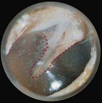
Figure 38-1 Tympanic membrane. Red dashed line, Handle of the malleus; green dotted line, light reflex.
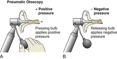
Note that with pneumatic otoscopy the normal ear drum moves in response to both positive and negative pressure gently applied with an insufflation bulb (Figure 38-2).
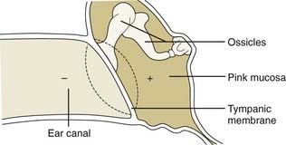
 Complete the dotted line to demonstrate movement of the normal tympanic membrane with positive pressure (Figure 38-3).
In AOM there is purulent middle ear effusion (MEE) and signs of inflammation that include otalgia or erythema of the TM. The TM is full and bulged outward, and there is no movement with insufflation (Figure 38-4).1 By contrast, in otitis media with effusion (OME), there is fluid in the middle ear space without signs of acute infection.2
 Complete the dashed and dotted lines demonstrating relative immobility in acute purulent otitis media. See Figure 38-5 for photographs of acute suppurative otitis media, otitis media with air-fluid level, and otitis media with effusion. Note that the distinction between and proper management of AOM and OME is discussed in Chapter 37, Ear Pain.
Stay updated, free articles. Join our Telegram channel

Full access? Get Clinical Tree


