T Lymphocytes, B Lymphocytes, and Natural Killer Cells

Chapter 117 T Lymphocytes, B Lymphocytes, and Natural Killer Cells


Defense against infectious agents is secured through a combination of anatomic physical barriers including the skin, mucous membranes, mucous blanket, and ciliated epithelial cells, as well as the various components of the immune system. The immune system of vertebrates integrates 2 fundamental response mechanisms. Innate (natural) immunity responds to infection regardless of previous exposure to the agent and includes polymorphonuclear leukocytes, dendritic and mononuclear phagocytic cells, various receptors that recognize common pathogen antigens (Toll-like receptors) and the complement system. Acquired (adaptive) immunity is a highly specific response that includes T lymphocytes, B lymphocytes, and natural killer (NK) cells. The immune system also helps protect against malignancy and autoimmunity.


Lymphopoiesis in the Fetus


Origin of the Lymphoid System


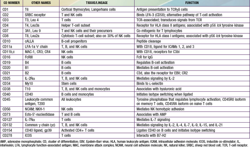
The human immune system arises in the embryo from gut-associated tissue. Pluripotential hematopoietic stem cells 1st appear in the yolk sac at 2.5-3 wk of gestational age, migrate to the fetal liver at 5 wk of gestation, and later reside in the bone marrow, where they remain throughout life (Fig. 117-1). Lymphoid stem cells develop from such precursor cells and differentiate into T, B, or NK cells, depending on the organs or tissues to which the stem cells traffic. Development of the primary lymphoid organs—thymus and bone marrow—begins during the middle of the 1st trimester of gestation and proceeds rapidly. Development of the secondary lymphoid organs—spleen, lymph nodes, tonsils, Peyer patches, and lamina propria—soon follows. These organs serve as sites of differentiation of T, B, and NK lymphocytes from stem cells throughout life. Both the initial organogenesis and the continued cell differentiation occur as a consequence of the interaction of a vast array of lymphocytic and microenvironmental cell surface molecules and proteins secreted by the involved cells. The complexity and number of lymphoid cell surface molecules led to the development of an international nomenclature for clusters of differentiation (CD) (Table 117-1).
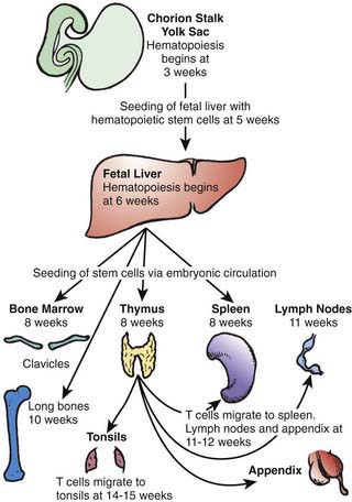


T and B lymphocytes are the only components of the immune system that have antigen-specific recognition capabilities and are responsible for adaptive immunity. NK cells are lymphocytes that are also derived from hematopoietic stem cells and are thought to have a role in host defense against viral infections, tumor surveillance, and immune regulation, but they do not have antigen receptors. Nonantibody proteins synthesized and secreted by T, B, and NK cells, and by the cells with which they interact, act as intercellular mediators and are referred to as cytokines or interleukins (ILs) (Table 117-2). Cytokines have the ability to act in an autocrine, paracrine, or endocrine manner to promote and facilitate differentiation and proliferation of the cells of the immune system.


Table 117-2 FUNCTIONAL CLASSIFICATION OF CYTOKINES*


2 Lymphocyte regulatory cytokines


GM-CSF, granulocyte macrophage colony stimulating factor; IFN, interferon; IL, interleukin; MHC, major histocompatibility complex; MCP-1, monocyte chemotactic protein; NK, natural killer; RANTES, regulated on activation normal t cell expressed and secreted; SCF, stem cell factor; TGF, transforming growth factor; TNF, tumor necrosis factor.


* This is not an exhaustive list.


Modified from Whiteside TL: Cytokine measurements and interpretation of cytokine assays in human disease, J Clin Immunol 14:327–339, 1994.


T-Cell Development and Differentiation


The primitive thymic rudiment is formed from the ectoderm of the 3rd branchial cleft and endoderm of the 3rd branchial pouch at 4 wk gestation. Beginning at 7-8 wk, the right and left rudiments move caudally and fuse in the midline. Blood-borne T-cell precursors from the fetal liver then begin to colonize the perithymic mesenchyme at 8 wk gestation. These precursor pro–T cells are identified by surface proteins designated as CD7 and CD34. At 8-8.5 wk gestation, CD7 cells are found intrathymically, with some cells that co-express CD4, a protein present on the surfaces of mature T-helper (TH) cells, or CD8, a protein found on both mature cytotoxic T cells and NK cells. In addition, some cells bear single T-cell receptor (Ti) chains (β, δ, or γ), but none bear complete T-cell receptors.


The mature T-cell receptor (TCR) is a heterodimer of 2 chains, either α and β or γ and δ, that is co-expressed on the cell surface with CD3, a complex of 5 polypeptide chains (γ, δ, ε, ζ, η). TCR gene rearrangement occurs by a process in which large, noncontiguous blocks of DNA are spliced together. These segments, known as V (variable), D (diversity), and J (joining), each have a number of variants. VDJ segments are joined to a constant region of the α gene, and VDJ segments are joined to the β gene to complete the receptor polypeptide genes. Random combinations of the segments account for much of the enormous diversity of TCRs that enables humans to recognize millions of different antigens. TCR gene rearrangement requires the presence of recombinase activating genes, RAG-1 and RAG-2, as well as other recombinase components. This process is flawed in mice with severe combined immunodeficiency (SCID) and in some humans with SCID. Rearrangement of TCR genes signifies commitment of pro–T cells to T-lineage development, becoming pre–T cells. TCR gene rearrangement begins shortly after colonization of the thymus with stem cells, and the establishment of the T-cell repertoire begins at 8-10 wk of gestation. By 9.5-10 wk, >95% of thymocytes express CD7, CD2, CD4, CD8, and c (cytoplasmic) CD3, and ≈30% bear the CD1 inner cortical thymocyte antigen. By 10 wk, 25% of thymocytes bear αβ TCRs. Ti αβ T cells gradually increase in number during embryonic life and represent >95% of thymocytes postnatally.


As immature cortical thymocytes begin to express TCRs, the processes of positive and negative selection take place. Positive selection

Stay updated, free articles. Join our Telegram channel

Jun 18, 2016 | Posted by in PEDIATRICS | Comments Off on T Lymphocytes, B Lymphocytes, and Natural Killer Cells

Full access? Get Clinical Tree

Get Clinical Tree app for offline access