25 Sports Medicine
This chapter provides a brief overview of several of the more common sports-related injuries.
Etiology and Pathogenesis
Pediatric athletes are anatomically and physiologically different from their adult counterparts. Developing bone is structurally weaker than adult bone. The physis, or growth plate, is between the metaphysis and epiphysis in the long bones of children. It is weaker than adjacent ligaments and thus more prone to fracture under high energy transfer. Physeal fractures are unique to children and account for 15% to 20% of all pediatric fractures (see Chapter 22).
Injuries by Body System
Shoulder Injuries
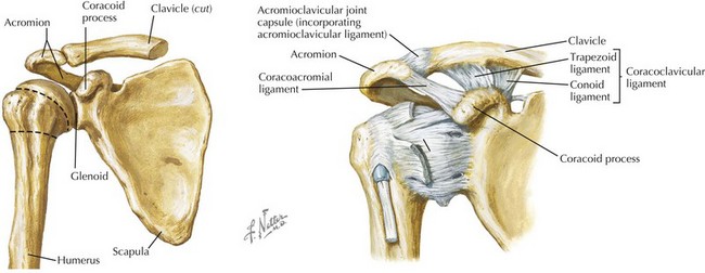
The shoulder girdle is composed of three joints: the sternoclavicular joint, acromioclavicular (AC) joint, and glenohumeral joint. The shoulder is the loosest joint in the body, with a shallow glenoid fossa and little bony support (Figure 25-1). Both acute traumatic and chronic overuse injuries are common.
Acromioclavicular Separation
The AC joint is composed of the acromion of the scapula and the distal clavicle. The AC and coracoclavicular ligaments stabilize the joint (see Figure 25-1). AC joint injuries, including sprains, subluxations and dislocations, are referred to as shoulder separations and account for 10% of all shoulder injuries. Injury to the AC joint is usually caused by a fall onto the top of the shoulder, causing the scapula and acromion to be pushed inferiorly, and the clavicle, with its medial end attached to the sternum, to be elevated. AC separations have a 5 : 1 male-to-female ratio and are encountered most frequently in hockey, wrestling, and the martial arts. Examination reveals asymmetric enlargement of the AC joint with localized tenderness to the joint. Forward flexion, extension, and cross-chest adduction results in AC joint pain. Diagnosis is made clinically or by plain radiographs dedicated to the AC joint. Milder separations are typically treated nonoperatively with rehabilitation, and the far less common severe separations may require surgical repair.
Anterior Shoulder Dislocation
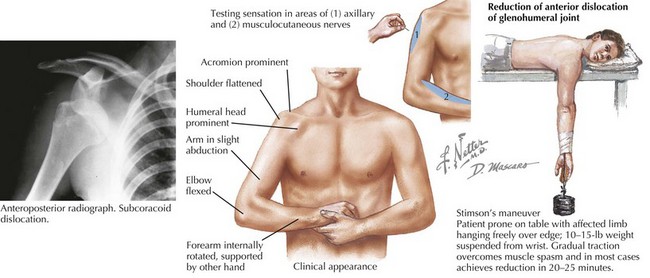
Examination reveals flattening of the deltoid prominence, prominence of the acromion, fullness of the subcoracoid region, and downward displacement of the axillary fold (Figure 25-2). It is also important to test for axillary and musculocutaneous nerve injury (see Figure 25-2).
Evaluation requires axillary, anteroposterior (AP), and scapular Y radiographs both before and after reduction to rule out associated fracture. An acute glenohumeral joint dislocation is an orthopedic emergency and requires closed reduction. There are many techniques for closed reduction; two of the more common are the traction–countertraction technique and Stimson’s maneuver (see Figure 25-2). It is important to reexamine the neurovascular integrity of the arm after reduction. Surgical reduction is rarely indicated. After reduction, the arm is immobilized for 2 to 4 weeks followed by gradual rehabilitation. Up to 90% of patients with shoulder dislocations before age 20 years have a recurrence.
Elbow Injuries
Elbow Dislocation
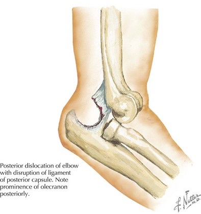
The elbow is the most commonly dislocated joint in children. Posterior dislocations are more common because of the shape of the olecranon process. They occur in older children, whose physes have closed, after falling backward onto an outstretched hand with the shoulder abducted. Simple dislocations can be seen in conjunction with associated fractures. On examination, an obvious deformity is noted, with the olecranon process displaced prominently behind the distal humerus (Figure 25-3). A careful neurovascular examination must be performed before and after reduction, paying particular attention to the median nerve. AP and lateral radiographs are useful in diagnosis. Isolated dislocation is treated by closed reduction. Unlike a shoulder dislocation, the greatest risk for an elbow dislocation is stiffness rather than recurrence. For this reason, immobilization is required for a short period of time, and return to full use is closely supervised.
< div class='tao-gold-member'>
Stay updated, free articles. Join our Telegram channel

Full access? Get Clinical Tree


