CHAPTER 29 Splenectomy
Step 1: Surgical Anatomy
♦ The spleen is positioned anterior to the costodiaphragmatic recess, lateral to the greater curvature of the stomach, adjacent to the tail of the pancreas, anterior to the left kidney, and superior or posterior to the splenic flexure of the colon.
♦ The parietal peritoneum adheres to the splenic capsule along its lateral border opposite the hilum. These attachments make up the suspensory ligaments of the spleen, which tend to be avascular.
♦ The splenorenal ligament extends from the left kidney to the hilum of the spleen and is where the splenic vessels and tail of the pancreas are invested.
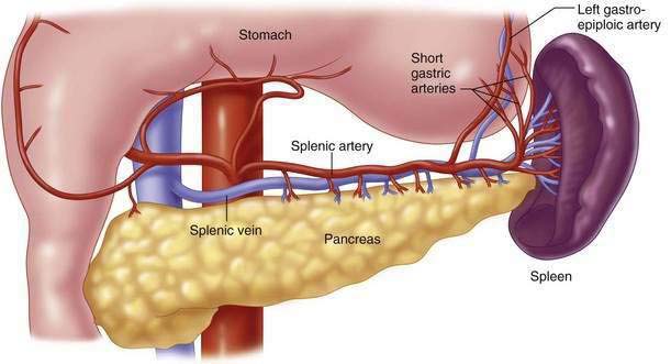
♦ The splenic artery arises from the celiac trunk and travels along the superior edge of the pancreas, where it branches to form the short gastric, left gastroepiploic, and terminal splenic branches (Fig. 29-1).
♦ The splenic vein forms in the splenorenal ligament and runs inferior to the artery and posterior to the pancreas. It joins the superior mesenteric vein behind the head of the pancreas to form the portal vein.
Step 2: Preoperative Conditions
Hematologic Disorders
Immune Thrombocytopenic Purpura
♦ Immune thrombocytopenic purpura (ITP) is characterized by a low platelet count, normal bone marrow, and an absence of other diseases that can cause thrombocytopenia.
♦ Splenectomy is used primarily only in patients with refractory severe symptomatic thrombocytopenia failing medical therapy.
 Indications include patients with ITP for 6 weeks and a platelet count of less than 10,000, irrespective of bleeding symptoms.
 Another indication is duration of ITP for 3 months and an incomplete response to primary therapy with a platelet count of less than 30,000.
 Indications include patients with ITP for 6 weeks and a platelet count of less than 10,000, irrespective of bleeding symptoms.
 Another indication is duration of ITP for 3 months and an incomplete response to primary therapy with a platelet count of less than 30,000.
Hereditary Spherocytosis
♦ An autosomal dominant disease resulting from a deficiency of spectrin, which leads to membrane abnormalities.
♦ Splenectomy is performed to correct the anemia, but removal of the spleen does not normalize the red blood cell morphology.
Sickle Cell Disease
♦ Elongated crescent-shaped erythrocytes are unable to deform within the microvasculature, resulting in thromboses and microinfarctions.
Tumors
♦ Splenectomy was formerly used to assist in the staging of Hodgkin’s lymphoma. However, with the increasing accuracy of noninvasive imaging, including computed tomography (CT) scan, lymphangiography, and positron emission tomography scans, the number of staging splenectomies performed has decreased.
Cysts
♦ True cysts are further subdivided into parasitic and nonparasitic cysts.


Stay updated, free articles. Join our Telegram channel

Full access? Get Clinical Tree


