CHAPTER 11 a nodule of normal splenic tissue commonly located near the splenic hilum. a decrease in hemoglobin levels in the blood. a rare benign neoplasm composed of lymphoid tissue. Also known as splenoma. the percentage of red blood cells in the blood. carries oxygen from the lungs to the cells and returns carbon dioxide back to the lungs. hematoma located within the splenic parenchyma. proliferation of white blood cells. white blood cell count above 20,000 mm3. white blood cell count below 4000 mm3. malignant disorder involving the lymphoreticular system. a localized dilatation of the splenic artery. occlusion of the main splenic artery or one of its branches. hematoma located between the splenic capsule and parenchyma. • Predominant organ in the left upper quadrant. • Except at the hilum, the spleen is covered by the peritoneum. • The spleen is divided into the: • The splenic artery arises from the celiac axis, courses along superior pancreatic borders, dividing into six branches after entering the splenic hilum. • The splenic vein joins the superior mesenteric vein, forming the main portal vein. • In cases of portal hypertension, the splenic vein may shunt blood directly into the left renal vein. • Predominantly located in the left hypochondriac region with the superior aspect extending into the epigastric region. • Located inferior to the diaphragm and anterior to the left kidney. • Lies posterior and lateral to the stomach. • No preparation is necessary for a sonogram of the spleen. • Nothing by mouth 6 to 8 hours before examination is the typical preparation, because imaging the spleen is rarely requested alone. • Use the highest-frequency abdominal transducer possible to obtain optimal resolution for penetration depth. • Place gain settings to display the normal splenic parenchyma as a medium shade of gray (similar to the liver) with adjustments to reduce echoes within the vessels. • Focal zone(s) at or below the place of interest. • Sufficient imaging depth to visualize structures immediately posterior to the region of interest. • Harmonic imaging or decreasing system compression (dynamic range) can be used to reduce artifactual echoes within anechoic structures. • Spatial compounding can be used to improve visualization of structures posterior to a highly attenuating structure. • Patients may lie in the supine, right posterior oblique, or right lateral decubitus position. • Coronal and transverse scanning planes are used to evaluate the spleen from the left hemidiaphragm to the left kidney. • Evaluation and documentation of the length, width, and thickness of the spleen. • Documentation and measurement of any abnormality in two scanning planes with and without color Doppler should be included. • Carries oxygen from the lungs to the tissues in the body. • Carries carbon dioxide back to the lungs. • Develops in the bone marrow and has a life span of 120 days. • Spleen stores red blood cells and destroys old red blood cells. • Elevation associated with polycythemia vera and severe diarrhea. • Decreases associated with internal bleeding, hemolytic anemia, Hodgkin’s disease, and hemangiosarcomas. • Oxygen-carrying pigment of the red blood cell. • Carries oxygen from the lungs to the cells and carbon dioxide from the cells back to the lungs. • Developed in the bone marrow inside the red blood cell. • Recycled by the spleen into iron.
Spleen
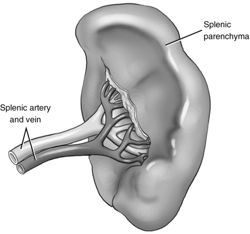
Anatomy (fig. 11-1)
Splenic vasculature
Location
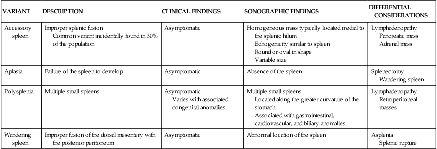
VARIANT
DESCRIPTION
CLINICAL FINDINGS
SONOGRAPHIC FINDINGS
DIFFERENTIAL CONSIDERATIONS
Accessory spleen
Improper splenic fusion
Common variant incidentally found in 30% of the population
Asymptomatic
Homogeneous mass typically located medial to the splenic hilum
Echogenicity similar to spleen
Round or oval in shape
Variable size
Lymphadenopathy
Pancreatic mass
Adrenal mass
Aplasia
Failure of the spleen to develop
Asymptomatic
Absence of the spleen
Splenectomy
Wandering spleen
Polysplenia
Multiple small spleens
Asymptomatic
Varies with associated congenital anomalies
Multiple small spleens
Located along the greater curvature of the stomach
Associated with gastrointestinal, cardiovascular, and biliary anomalies
Lymphadenopathy
Retroperitoneal masses
Wandering spleen
Improper fusion of the dorsal mesentery with the posterior peritoneum
Asymptomatic
Abnormal location of the spleen
Asplenia
Splenic rupture
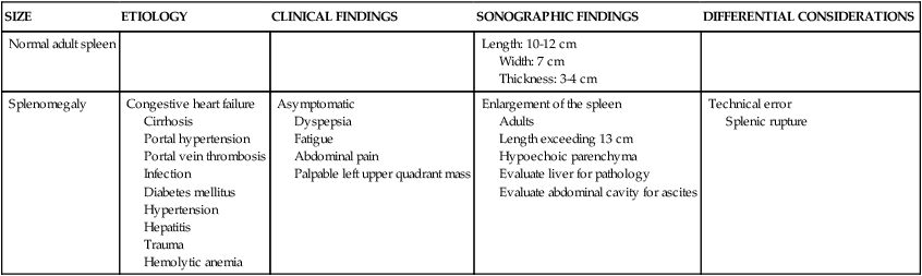
SIZE
ETIOLOGY
CLINICAL FINDINGS
SONOGRAPHIC FINDINGS
DIFFERENTIAL CONSIDERATIONS
Normal adult spleen
Length: 10-12 cm
Width: 7 cm
Thickness: 3-4 cm
Splenomegaly
Congestive heart failure
Cirrhosis
Portal hypertension
Portal vein thrombosis
Infection
Diabetes mellitus
Hypertension
Hepatitis
Trauma
Hemolytic anemia
Asymptomatic
Dyspepsia
Fatigue
Abdominal pain
Palpable left upper quadrant mass
Enlargement of the spleen
Adults
Length exceeding 13 cm
Hypoechoic parenchyma
Evaluate liver for pathology
Evaluate abdominal cavity for ascites
Technical error
Splenic rupture
Technique
Preparation
Examination technique and image optimization
Laboratory values
Erythrocyte
Hemoglobin
PATHOLOGY
ETIOLOGY
CLINICAL FINDINGS
SONOGRAPHIC FINDINGS
DIFFERENTIAL CONSIDERATIONS
Abscess
Infective endocarditis—most common
Infection
Trauma
Fever
Left upper quadrant (LUQ) pain
Leukocytosis![]()
Stay updated, free articles. Join our Telegram channel

Full access? Get Clinical Tree

 Get Clinical Tree app for offline access