and Marcelo Zugaib4
(1)
São Paulo University, Bauru, Brazil
(2)
Parisian University, Bauru, France
(3)
Member of International Fetal Medicine and Surgery Society, Bauru, Brazil
(4)
Obstetrics, University of São Paulo, Bauru, Brazil
Fetal evaluation from its cranial-most portions, in the first trimester, gives us a unique opportunity to track abnormalities, through the measurement of nuchal translucency (a simple fluid accumulation, without septa, in the posterior region of the fetal occiput). This is performed in an ideal period ranging from 11 weeks to 13 weeks and 6 days of pregnancy (CRL measuring 45–84 mm). An increase in the fluid in this region correlates with chromosomal abnormalities, genetic diseases, and heart defects.
Cystic hygroma, a large cystic area, sometimes septate, in the same topography (posterior region of the fetal occipital bone), may assume large proportions, being common in cases of fetal Turner syndrome (karyotype = 45,X0). It is also a marker of genetic diseases. Cystic hygroma associated with Turner syndrome is rarely observed in late stages of pregnancy because of the high rates of miscarriages observed in these cases. In survivors, a complete resorption of liquid occurs until the end of pregnancy.
At more advanced stages of pregnancy (second and third trimesters) when a cystic formation in the cervical region is detected, it is necessary to consider a lymphangioma as a differential diagnosis. This type of lesion often has less symmetrical distribution, a more heterogeneous aspect than a hygroma and may also occur in other parts of the fetal body, such as the underarm regions and the sacral region.
The accumulation of widespread liquid, involving subcutaneous tissue and cavity effusion (pleural, abdominal, pericardial), is called hydrops, and this condition may be a consequence of immune disorder. In many cases, no cause is found for this form of edema. Some authors consider that the term hydrops should only be used in cases of liquid spills in at least two cavities or stroke in a cavity accompanied by edema of at least 5 mm in subcutaneous tissue. Often, this medical condition is accompanied by placental thickening.
Tumors in the fetal head can affect the central nervous system and shallow portions, such as the skin and subcutaneous tissue. Congenital tumors affecting the brain are quite infrequent. Cystic formations due to fetal tumors are discussed in the relevant chapter (see Chap. 3 on the Central Nervous System). Solid or cystic tumors are rarely seen and are mostly represented by teratomas. Although they consist of histologically benign tumors, devastating consequences for the fetus are sometimes observed. Its growth may be limited to the brain or even exceed the limits of the skull.
Visceral tumors of the lungs, heart, and abdominal organs are discussed in their respective chapters.
The most frequent solid tumors of the fetal face are hemangiomas.
In the cervical region, in addition to hemangiomas, other conditions may be observed, such as goiter, branchial cysts (always with a good prognosis), and teratomas.
Toward the caudal regions, the fetal tumors that are most commonly diagnosed are sacrococcygeal teratomas. These may be small or minimal, but they can achieve giant dimensions. These tumors affect caudal surface portions or invade the pelvis and abdomen of the fetus. The most aggressive tumors often bring drastic consequences, as a result of blood sequestration, and may cause fetal death.


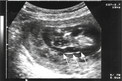
Fig. 13.1
Longitudinal view of the fetus at 13 weeks showing increased nuchal translucency (arrows)

Fig. 13.2
Increased nuchal translucency

Fig. 13.3
Sagittal section (right) and transverse section (left) demonstrating increased nuchal translucency (arrows)

Fig. 13.4
Sagittal section of an embryo at 8 weeks and 4 days with increased fluid, extending from the nuchal region to the caudal region (arrows)

Fig. 13.5
Sagittal section of a 16-week fetus demonstrating an extensive cervical cystic hygroma, accompanied by ascites. Col = column, Higr = hygroma. Asc = ascites

Fig. 13.6
Cross-section of a 16-week fetal head, demonstrating a chambered cystic hygroma

Fig. 13.7
A similar case of Fig. 13.6 now with a zoomed image showing cystic hygroma (HIG). Polo cef = fetal head

Fig. 13.8
Sagittal section of 13-week fetus with Turner syndrome (45,X0) showing increased nuchal translucency (arrows) and fluid accumulation under the scalp

Fig. 13.9
Increased nuchal translucency (arrows and calipers) in the context of genetic disease (first-degree cousins as parents)

Fig. 13.10
Cross-section of a 13-week fetal head, normal karyotype, and increased fluid in the neck region (arrows)

Fig. 13.11
Cross-section in a cephalic pole (PC) showing cystic hygroma (H), with the typical Y shape of the septa (S)

Fig. 13.12
Skin excess in the neck region (arrows), following the cystic hygroma. The karyotype revealed Turner syndrome (45,X0)

Fig. 13.13
Coronal section of a 15-week fetus presenting extensive lymphangioma (arrows), extending from the cephalic pole (PC) to the body. Note the presence of large cysts (L) and the heterogeneous aspect of the lesion

Fig. 13.14
Extensive lymphangioma. L = lymphatic cysts

Fig. 13.15
Cross-section of a fetal head showing a heterogeneous mixed mass continuous with the brain parenchyma, corresponding to teratoma with exophytic growth (arrows)


