Fig. 1.1
Location of primary tumors in osteosarcoma and Ewing sarcoma (percentage)
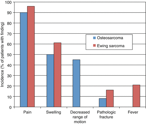
Fig. 1.2
Presenting symptoms in osteosarcoma and Ewing sarcoma (percentage)
1.3 Sex and Ethnicity
There is a slight male predominance in malignant bone tumors overall, 1.19:1 (male to female) (1999), which also is true for each histologic subgroup: 1.27:1 for Ewing, 1.16:1 for osteosarcoma, and 1.5:1 for chondrosarcoma. These differences become even more pronounced in the adolescent age group (15–19 years old) with 2.03:1, 1.92:1, and 1.71:1 male to female predominance for Ewing, osteosarcoma, and chondrosarcoma, respectively (2006; Stiller et al. 2006).
A striking difference in incidence of Ewing sarcoma has been demonstrated between the white non-Hispanics and Hispanics groups and the African American/Black and Asian/Pacific Islander groups. Ewing sarcoma is extremely rare in groups of African and Southeast Asian descent; in fact, a SEER report demonstrated no cases of EWS in adolescents and young adults in these ethnic groups (2006). In osteosarcoma the ethnic predilection is reversed and slightly more common in African American children, but the difference between the two is much smaller (1:1.33; white to black, respectively) (1999). Given the higher overall incidence of osteosarcoma than Ewing sarcoma, the total bone tumor ethnic ratio between white and black is 1.3:1 (1999).
1.4 Sites of Tumors
Osteosarcoma most commonly occurs in the long bones of the lower limb, while Ewing sarcoma and chondrosarcoma more commonly occur in the axial skeleton as well as long bones (1999; 2006). More specifically, osteosarcoma generally presents in the metaphyseal region of long bones, with the distal femur, proximal tibia, and proximal humerus being the most common sites in order of frequency. Ewing sarcoma is more likely to be seen in the diaphyseal regions of long bones and flat bones, with 40 % of Ewing tumors occurring in the axial skeleton (Arndt and Crist 1999). Non-skeletal presentations occur within the Ewing sarcoma family of tumors (ESFT) as well and generally demonstrate the same histologic findings as well as the characteristic EWS/FLI1 translocation. Askin tumors are ESFTs that involve the chest wall and clearly present a site-specific local control challenge (Shamberger et al. 2003). Ewing sarcoma also occurs in extraosseous sites, and while they were originally treated according to rhabdomyosarcoma chemotherapy regimens, patients with this subtype of the ESFT are now treated using Ewing sarcoma-specific regimens (Zagar et al. 2008; Raney et al. 1997). Such extraosseous tumors may occur in the soft tissue, retroperitoneum, skin, and solid organs such as the kidney (Orr et al. 2012; Zollner et al. 2013).
1.5 Signs and Symptoms
Pain and swelling are the primary presenting features for both osteosarcoma and Ewing sarcoma. Approximately 70 % of patients present with pain, and about 20 % have a concomitant palpable mass (Widhe and Widhe 2000). In a report from the University of Sao Paolo, investigators described that almost 90 % of patients had pain, but pain at rest was noted in less than 10 % of patients. A tumor mass was noted in 57 % of patients (Guerra et al. 2006). Night time pain, classically thought to be an indicator of malignancy, only occurs in 20 % of patients with bone sarcoma (Widhe and Widhe 2000), and preceding trauma, which can be diagnostically misleading, was reported in almost 50 % of patients with osteosarcoma. Patients with extraosseous Ewing sarcoma typically present features with pain and swelling in two-thirds of patients. Rare presentations in this group include shortness of breath, difficulty walking, urinary obstruction, or constipation (Orr et al. 2012). Constitutional symptoms including fever, weight loss, fatigue, and loss of appetite occurred in 31 % of patients with skeletal Ewing sarcoma and 20 % of patients with extraosseous Ewing sarcoma and are more frequent in patients with metastatic disease (Biswas et al. 2014). Patients with Ewing sarcoma of the paraspinal region may develop epidural invasion and present with signs of neurologic compromise from spinal cord or nerve root compression, including paralysis and bowel or bladder dysfunction. Such patients require emergent evaluation and laminectomy for neurological recovery. Patients with pelvic and chest wall tumors may not present with a palpable mass due to the fact that the growth of the tumor is likely internal or intracavity. Consequently patients with chest wall tumors may present with cardiorespiratory symptoms if there is significant lung compression and mediastinal shift. Both osteosarcoma and Ewing sarcoma can also metastasize to the lungs and skeletal bones, although metastatic lung nodules are rarely large enough at presentation to cause respiratory symptoms. Patients may also present with a pathological fracture at time of initial diagnosis. Plain radiographs were acquired on almost two-thirds of patients reported by Widhe et al. (Widhe and Widhe 2000), and many of these were done to exclude the trauma/fracture. Only 2 patients out of 100 with osteosarcoma were found to actually have a pathological fracture at diagnosis, and no patients out of the 47 with Ewing sarcoma were found to have a fracture. The typical differential diagnosis for patients who present as described above includes tendinitis, tumor, or trauma, and more uncommonly bursitis, Osgood-Schlatter disease, or Legg-Calve-Perthes disease, among many others (Widhe and Widhe 2000).
Patients with osteosarcoma have an average delay of 15 weeks from development of symptoms to diagnosis, while delays of up to 34 weeks have been reported for patients with Ewing sarcoma (Widhe and Widhe 2000; Sneppen and Hansen 1984; Goyal et al. 2004). The older age distribution for malignant bone tumors, the typical denial of symptoms in adolescence, and the frequent challenges in delineating a malignant lesion from a simple trauma, infection, or other inflammatory condition likely contribute to delays in the recognition of symptoms and diagnosis (Widhe and Widhe 2000; Goyal et al. 2004; Sneppen and Hansen 1984). Initial recommendations of simple rest and analgesics may also prolong time to diagnosis since temporary improvements in pain and tenderness are frequently seen with such measures and may reinforce the impression of benign etiology such as tendinitis (Widhe and Widhe 2000). Reassuringly, the data suggest that such delays do not result in adverse survival outcome for patients (Brasme et al. 2014; Martin et al. 2007).
1.6 Diagnostic Evaluation
Malignant bone tumors are rare, and there is a broad differential for bone pain especially in the pediatric and adolescent age group; therefore, the suspicion of such tumors by primary care physicians is low. Suspicion for malignancy at first medical visit has been noted for only 31 % of patients diagnosed with osteosarcoma and 19 % of patients with Ewing sarcoma (Widhe and Widhe 2000). Conventional imaging features with plain x-ray help differentiate benign bone lesions from malignant disease. Typical features of malignancy including cortical bone disruption, periosteal elevation, subperiosteal new bone formation, lytic and sclerotic changes in bone, soft tissue mass, and soft tissue ossification (classical sunburst appearance with malignant osteosarcoma), which is discussed in detail in Chap. 2 (Murphey et al. 1997). Widhe et al. reported that 67 % and 60 % of patients ultimately diagnosed with osteosarcoma and Ewing sarcoma, respectively, had x-ray imaging performed after their first primary care visit (Widhe and Widhe 2000). Unfortunately the false-negative rate, at this initial image, was 9 % for osteosarcoma and 43 % for Ewing sarcoma. Other imaging required for the evaluation of a newly diagnosed patient with a primary bone malignancy includes MRI of the primary tumor to determine osseous, joint, and soft tissue tumor extent, along with other important characteristics for surgical planning such as neurovascular encroachment. Also required is a CT of the chest to evaluate for parenchymal metastatic lung disease and a technetium bone scan or CT-PET for distant skeletal and bone marrow disease (Meyer et al. 2008; Franzius et al. 2000). Radiological features typical for osteosarcoma (sunburst appearance, metaphyseal lesion) and Ewing sarcoma (onion skin appearance to periosteal elevation and diaphyseal lesion) can be considered in addition to clinical features such as age and ethnicity to predict tumor type. However, while sensitive, these features are not specific and a histological diagnosis is critical for a correct diagnosis and treatment planning. Laboratory evaluation of tumor markers such as alkaline phosphatase and LDH also provide some prognostic information (Bielack et al. 2002). Bone marrow aspirate and biopsy are still conventionally required to stage patients with Ewing sarcoma, but may soon be replaced with CT-PET which is very sensitive to presence of bone marrow disease (Hawkins et al. 2005; Newman et al. 2013).
Stay updated, free articles. Join our Telegram channel

Full access? Get Clinical Tree


