Fig. 20.1
Variations of the unicorneate uterus: a communicating, b noncommunicating rudimentary horn, c rudimentary horn without a cavity, and d isolated unicorneate uterus [1]

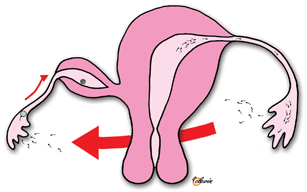
Fig. 20.2
Pregnancy in a noncommunicating rudimentary uterine horn arising due to transperitoneal migration of sperm
In a review of 568 cases of rudimentary uterine horn pregnancies from 1900 to 1999, a rupture rate of 50 % was found with 80 % occurring before the third trimester [7]. The maternal mortality rate has fallen dramatically over time, from 23 % in the 1920s to currently less than 0.5 % as a result of improved diagnosis and early treatment. Only a few reports exist of rudimentary horn pregnancies proceeding to term [8].
Women with uterine malformation have an increased risk of miscarriages and obstetric complications including intrauterine growth retardation, abnormal presentations, and preterm labor [9]. It is possible that uterine anomaly contributes to the preterm labor in our patient’s first pregnancy. Operative record of the cesarean delivery stated “a fibroma-like structure at the departure of the left salpinx.” Uterine anomalies can be classified according to the guidelines developed by the American Society of Reproductive Medicine [10], or according to a recent classification system for female genital anomalies developed by the European Society of Human Reproduction and Endocrinology (ESHRE) and European Society for Gynecological Endoscopy (ESGE) [4].
Management
When a pregnant women presents with abdominal pain and symptoms of hemorrhagic shock , immediate surgical intervention should be performed. The choice of operative method depends on the condition of the patient and surgeon’s expertise and experience. Laparoscopy may be an alternative to laparotomy in the case of a hemodynamically stable patient. Surgery should include excision of the entire rudimentary horn. In addition to the morbidity associated with uterine rupture , a rudimentary horn pregnancy carries an increased risk of abnormal implantation of the placenta, such as placenta accreta or percreta. Thus, the clinician should be prepared for the potential massive intraabdominal hemorrhage.
Early detection of rudimentary uterine horn pregnancies remains a clinical challenge. Even though more women undergo a first trimester ultrasound examination, the sensitivity of ultrasound in diagnosing rudimentary horn pregnancies is only 29 % [2]. Moreover, the diagnosis may be masked by a history of previous normal pregnancies, as in this case. On pelvic examination, the patient may have an adnexal mass, causing deviation of the uterus to one side. Ultrasound findings may be suggestive of the presence of a bicornuate uterus.
The rudimentary horn pregnancy may be detected by the lack of continuity between the cervical canal and the lumen of the uterine horn. In some cases, a well-defined placenta fitting into the confines of the rudimentary horn may differentiate a rudimentary horn pregnancy from an abdominal pregnancy [6]. The diagnosis may be confirmed by magnetic resonance imaging. Once the diagnosis of rudimentary horn pregnancy is established, surgery should be performed including removal of the ipsilateral Fallopian tube to prevent future ectopic pregnancies [2]. In rare cases, expectant management may be attempted [7], but this requires close monitoring of fetal growth and myometrial thickness and access for immediate operative intervention. If a rudimentary uterine horn is identified before conception, elective surgical removal by laparoscopy is recommended [3].
Stay updated, free articles. Join our Telegram channel

Full access? Get Clinical Tree


