1 Resuscitation
Cardiopulmonary resuscitation (CPR) is the series of emergency interventions provided to a person who appears dead or in respiratory extremis, with the goal of restoring vital functions through optimization of cardiac output and tissue oxygen delivery. The two main components are external cardiac massage (chest compressions) and assisted respirations.
Most children who require CPR do not survive. Those who do survive often have significant neurologic deficits from the hypoxia and ischemia associated with the cardiopulmonary arrest. However, some children do return to their premorbid function. This may be related to recognition and treatment of impending cardiorespiratory failure, early bystander CPR, or rapid correction of the life-threatening event. It is difficult to predict which patients will have a return of spontaneous circulation and ultimately survive. Therefore, high-quality CPR should be begun immediately while more information is gathered to guide therapy.
The incidence of out-of-hospital cardiopulmonary arrest is difficult to ascertain from the current literature. However, data from multicenter registries support many widely held beliefs about pediatric out-of-hospital arrests. The incidence of non-traumatic, out-of-hospital cardiac arrest is highest for infants (children younger than 1 year of age). The most common cause in this age group is sudden infant death syndrome (SIDS). The presenting rhythm is usually asystole, and survival to discharge is rare (≈3%). For all other pediatric age groups, trauma is the leading cause of death. Although asystole is the most common arrest rhythm, ventricular tachycardia (VT) and ventricular fibrillation (VF) do occur and are more common in older children, especially adolescents. Survival in patients with ventricular dysrhythmias is higher than in patients with cardiopulmonary arrest associated with rhythms that are not responsive to cardioversion or defibrillation, such as asystole and pulseless electrical activity (PEA). For nontraumatic arrests, survival even for older children remains low, about 9% in those older than 1 year of age.
Etiology and Pathogenesis
Cardiopulmonary arrest may result from many causes. Sepsis, respiratory infection, pulmonary conditions, drowning, SIDS, and injuries can lead to respiratory failure or shock. Without effective intervention, cardiopulmonary failure (inadequate perfusion and ineffective or absent respiration) ensues. Most pediatric arrests occur after an initial respiratory arrest (rather than circulatory failure) and if prolonged result in terminal rhythms of bradycardia; PEA; and, finally, asystole. Patients in asystole likely have experienced a significant hypoxic–ischemic insult.
Clinical Presentation
The signs and symptoms of children requiring immediate resuscitation are typically the result of failure of the delivery of two vital substrates—oxygen and glucose—to end organs (Table 1-1). Recognition of these manifestations through a physical examination that focuses on airway, gas exchange, and cardiovascular stability allows for rapid resuscitation of those who have failure of substrate delivery and identification of those at risk for failure.
Table 1-1 Signs of a Life-Threatening Condition
Airway | Complete or severe airway obstruction |
Breathing | Apnea, significant work of breathing, bradypnea |
Circulation | Absence of detectable pulses, poor perfusion, hypotension, bradycardia |
Disability | Unresponsiveness, depressed consciousness |
Exposure | Significant hypothermia, significant bleeding, petechiae or purpura consistent with septic shock, abdominal distension consistent with acute abdomen |
Adapted from American Academy of Pediatrics and American Heart Association: Pediatric Advanced Life Support. Dallas, TX, American Heart Association Publication, 2006.
Initial Assessment
Evaluation of a critically ill or injured child should begin with a general assessment. Physical examination clues help the provider determine the extent of illness or injury (i.e., whether the condition is life threatening or not) and identify systems that require closer attention during the remainder of the assessment. The Pediatric Assessment Triangle of the Pediatric Advanced Life Support (PALS) course outlines the following components of the general assessment:
The initial assessment can be done without laying hands on the patient and should take no more than several seconds. If the patient’s condition is life threatening, additional support should be recruited immediately. After these rapid initial impressions, the clinician should aim to perform a swift yet careful primary assessment.
Primary Assessement
The primary assessment evaluates and addresses vital functions in a systematic way with priority to systems that are most crucial for sustaining life. Conveniently, the components of the primary assessment can be remembered as the “ABCDEs”—airway, breathing, circulation, disability, and exposure and environment. If a life-threatening abnormality is identified at any point, the aberration should be addressed before moving on in the assessment.
With the publication of new guidelines for cardiopulmonary resuscitation in 2010, it has been recommended that the standard sequence of “ABC” be switched to “CAB” for patients who need cardiopulmonary resuscitation. This recommendation is based on the recognition that most cardiopulmonary arrests occur in adults and that even in children and adolescents sudden arrest is more likely to be due to a cardiac arrhythmia. Prompt institution of cardiac compressions to provide artificial circulation is important to ensure the best possible outcome for individual patients. This is especially important in out-of-hospital settings where early initiation of bystander CPR has been shown to be one of the strongest predictors of survival and good neurologic outcome. In medical settings the approach to resuscitation may be individualized based on the clinical scenario. Close attention to securing the airway and providing artificial ventilation remain important but in the setting of cardiopulmonary arrest chest compressions should not be delayed.
Airway
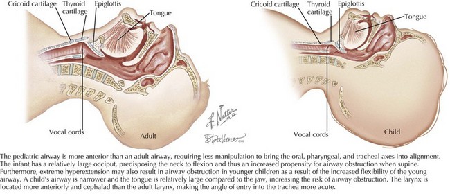
The patient’s airway is the first priority. There are fundamental differences between the airway of a child and that of an adult. The pediatric airway (Figure 1-1) is more anterior than the adult airway, requiring less manipulation to bring the oral, pharyngeal, and tracheal axes into alignment. In addition, the head-to-body proportion is larger in infants than in adults, and thus extreme hyperextension of the neck may exacerbate airway obstruction in younger children. The pediatric airway is narrower, and the tongue is relatively large compared with the jaw, increasing the risk of airway obstruction. The pediatric larynx is located more anteriorly and cephalad than the adult larynx.
The provider should assess airway patency using the “look, listen, and feel” approach. The provider should look at the chest wall and listen to the mouth and nose to detect whether there is evidence of air movement. Findings suggesting airway obstruction include increased respiratory effort with retractions, abnormal inspiratory sounds, or episodes during which no airway or breath sounds are produced despite respiratory effort. If a child is speaking, crying, or otherwise verbalizing, the airway is intact. Attention should be paid to the quality of the sounds. A hoarse or high-pitched cry should alert the provider to the possibility of airway compromise without complete obstruction. Finally, the provider should feel for air movement using a hand or cheek close to the patient’s mouth.
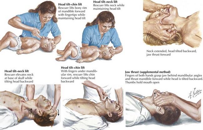
The most effective maneuvers for opening an obstructed pediatric airway are the head tilt–chin lift or jaw thrust techniques (Figure 1-2). In the head tilt–chin lift maneuver, the head is tilted back slightly (without overextending), and the chin is lifted gently with one finger on the bony prominence to avoid placing pressure on the soft tissues of the neck. A roll or towel may be placed under the shoulders to maintain the position.
If there is a risk of a neck injury, it is critical to stabilize the cervical spine during evaluation of the airway and avoid extending the neck. Manual cervical spine stabilization is accomplished by holding the head in the midline position while applying gentle cephalad traction. A cervical collar may be applied, taking care to size the collar appropriately. It is important to remember that neither manual nor collared stabilization provides true immobilization.
The jaw thrust maneuver should be attempted in these cases by lifting the jaw forward with the provider’s third or fourth fingers (or both) “hooked” under the angles of the mandible while avoiding compression of the soft tissues. The goal is to pull the mandibular block of tissue forward so that the lower central incisors are anterior to the upper central incisors.
At any point, if it is determined that the patient is unable to independently maintain the patency of his or her airway, the provider should open the airway to maintain adequate ventilation and protection from the aspiration of stomach contents. Simple suctioning should be attempted first because it may relieve an airway obstructed by secretions or foreign materials. Bag–valve–mask (BVM) ventilation (see below) may provide an open airway if a good mask seal is achieved and airway positioning is maintained.
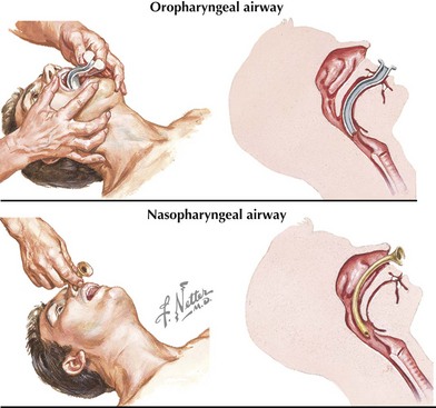
In an unconscious patient, an oropharyngeal airway can be used to help stent the mandibular block of tissue away from the posterior hypopharynx (Figure 1-3). A nasopharyngeal (NP) airway is another option. NP airways are well tolerated in unconscious and semiconscious patients and may even be used in conscious individuals with upper airway obstruction. NP airways should be used with caution when midface trauma is suspected because of the risk of inserting the airway through fractured bone into intracranial structures. Laryngeal mask airways (LMAs) are supraglottic airway devices that are being increasingly used in resuscitation settings to help bypass the soft tissues of the anterior oropharynx and to deliver oxygen directly to the proximal trachea.

Stay updated, free articles. Join our Telegram channel

Full access? Get Clinical Tree

