Chapter 73 Respiratory Distress Syndrome (Case 32)
Patient Care
Clinical Thinking
• Support airway and spontaneous breathing effort, providing supplemental oxygen and ventilatory support as needed.
History
• Are there risk factors for infection, such as maternal fever, untreated maternal group B streptococcus (GBS) colonization, prolonged rupture of membranes, and signs of fetal distress?
• Was meconium-stained amniotic fluid present? Were there signs of fetal hypoxemia or stress in utero (abnormal fetal heart tracings, abnormal umbilical artery blood gas levels)?
Physical Examination
• Vital signs: Abnormal respiratory pattern or effort may be present. Pulse oximetry may reveal hemoglobin desaturation. Bradycardia is commonly seen in the presence of hypoxia. Hypothermia itself may lead to respiratory distress. Tension pneumothorax results in hypotension.
• Signs of respiratory distress such a nasal flaring, grunting, or intercostal or subcostal retractions are often present.
• Hypotonia is often seen in sepsis, severe hypoxia, neurologic disorders, or exposure to maternal medications/drug use.
• A cardiac murmur, enlarged liver, or poor peripheral pulses are present in some congenital heart defects.
• Passage of meconium in utero often leads to staining of the finger nails or umbilical cord and meconium at or below the vocal cords.
Clinical Entities: Medical Knowledge
| Respiratory Distress Syndrome | |
|---|---|
| Pϕ | RDS results from a deficiency of pulmonary surfactant. Surfactant, composed of phospholipids and proteins, reduces alveolar surface tension. Surfactant production by type 2 pneumocytes is generally sufficient by 34 to 35 weeks’ of gestation. Inadequate surfactant production often leads to alveolar collapse. Primarily a disease of preterm infants, RDS may present in term infants with delayed lung maturity or decreased surfactant production (infants of diabetic mothers, perinatal asphyxia), or when surfactant is inactivated (meconium aspiration). Antenatal administration of glucocorticoids to mothers at risk for preterm delivery also accelerates lung maturity and has been proven to reduce the risk for RDS. |
| TP | Symptoms include nasal flaring, tachypnea, cyanosis, retractions, and expiratory grunting. Infants present with distress within the first 6 hours of life. The clinical course is characterized by progressive worsening of symptoms with a peak severity at 48 to 72 hours. |
| Dx | The diagnosis of RDS is based upon the combination of history, clinical features, and characteristic chest radiographs. Decreased breath sounds may be present. Chest films typically reveal a homogeneous “ground glass” or reticulogranular appearance secondary to diffuse atelectasis throughout the lung fields, and air bronchograms. ABG reveals hypoxemia, hypercarbia, and mixed acidosis. |
| Tx | Surfactant replacement therapy is either given prophylactically in the delivery room to at-risk infants or as a rescue therapy after clinical decompensation. Nasal CPAP helps to maintain functional residual capacity (FRC) and prevent alveolar collapse. Some infants require intubation and mechanical ventilation. See Nelson Essentials 61. |
| Transient Tachypnea of the Newborn | |
|---|---|
| Pϕ | This respiratory distress is caused by transient pulmonary edema secondary to delayed reabsorption of fetal lung fluid. Early in gestation, the pulmonary epithelium contributes to lung development and alveolar formation by secreting chloride into the alveolar space, creating an osmotic gradient that induces the flow of liquid in the air spaces. As the fetus matures, the pulmonary epithelium develops the capacity to reabsorb sodium. The surge of endogenous steroids and circulating catecholamines during labor and delivery further stimulates these epithelial cells to reabsorb sodium, pumping it into the interstitial space. Fetal lung fluid follows. Furthermore, prostaglandin secretion during labor leads to lymphatic dilatation, which aids in the clearance of fluid from the interstitium. |
| TP | Nasal flaring, retractions, grunting, tachypnea and cyanosis are often seen in the first few hours of life. When mild, TTN may last 12 to 24 hours. More severe cases may last 72 hours or longer. |
| Dx | TTN is a diagnosis of exclusion. Chest radiographs often reveal prominent perihilar streaking as a result of the engorgement of the lymphatic system and may have visible fluid in the fissures. ABG may reveal hypoxemia. |
| Tx | This is a self-limiting disease, and treatment is supportive. Supplemental oxygen or nasal CPAP is the mainstay of therapy. Infants may require IV fluids and gavage feeding until symptoms resolve. (See Chapter 66, Transient Tachypnea of the Newborn.) See Nelson Essentials 61. |
| Infection/Pneumonia | |
|---|---|
| Pϕ | Respiratory distress is often the first sign of infection in the neonate, and the lungs represent a common site for the establishment of sepsis in the newborn. Infections in the newborn may be bacterial or viral and may be acquired prenatally, during delivery, or early in the postnatal course. Most common causative agents are GBS, Escherichia coli, Listeria monocytogenes, and nontypable Haemophilus influenzae. Risk factors include prolonged rupture of membranes, chorioamnionitis, and fetal gasping due to asphyxia, which leads to increased risk for aspiration. |
| TP | These infants are typically lethargic and have some degree of respiratory distress. They may have trouble maintaining their body temperature. Eventually these infants demonstrate hypotension and poor perfusion as a sign of shock. |
| Dx | Chest radiographs often reveal patchy infiltrates, but they may also look similar to those seen in RDS, especially in the case of GBS pneumonia. White blood cell count, differential, platelet count, blood cultures, and Gram stain may be helpful. A spinal tap is necessary if the clinical suspicion for sepsis if high. Tracheal cultures should be interpreted with caution because it may not be possible to differentiate between true infection and early bacterial colonization. |
| Tx | Empiric use of intravenous antibiotics at the onset of distress to cover the most common pathogens and respiratory support are the mainstay of therapy. See Nelson Essentials 110. |
| Pneumothorax | |
|---|---|
| Pϕ | Pneumothorax is accumulation of free air from ruptured alveoli between the parietal pleural lining of the chest wall and the visceral pleura of the lung. A tension pneumothorax occurs when the trapped air impedes venous return to the heart, leading to decreased cardiac output and hemodynamic instability. |
| TP | An air leak should be suspected in any infant who has a sudden onset of respiratory distress or whose condition suddenly deteriorates. Infants often present with grunting, increased pallor or cyanosis, and retractions. |
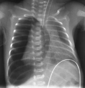
| Dx | If there is a unilateral pneumothorax, breath sounds may be decreased on the affected side. Transillumination of the chest wall with a fiberoptic light source may be extraordinarily helpful, because intervention may be necessary before a chest film is obtained. Chest radiographs typically reveal a sharply demarcated, hyperlucent area without pulmonary parenchymal markings (Figure 73-1). Lateral decubitus films, in which the affected side is positioned up, are quite helpful. Hypoxemia on ABG and sudden drop in oxygen saturations on pulse oximetry are characteristic findings. |
| Tx | No specific treatment is necessary for an asymptomatic pneumothorax. Urgent chest tube insertion into the pleural space, anterior to the lung, is the definitive treatment for symptomatic infants. See Nelson Essentials 138. |
< div class='tao-gold-member'>
Only gold members can continue reading. Log In or Register to continue
Stay updated, free articles. Join our Telegram channel

Full access? Get Clinical Tree


