Renal Neoplasms

59 Renal Neoplasms


Renal tumors account for 6% of all childhood cancers and are the sixth most common form of cancer in children. As with many pediatric cancers, there is no known environmental cause for childhood kidney tumors. Certain genetic syndromes are associated with a predisposition to developing renal neoplasms, specifically Wilms’ tumor. The etiology of many renal tumors may be attributable to tumor-specific chromosomal abnormalities, discussed below. Although Wilms’ tumor is one of the most curable tumors of childhood, other more rare pediatric renal tumors have a high mortality rate. Therefore, optimal management of the more rare renal tumors, such as tailoring therapy to tumor-specific genetic alterations to improve cure rates, is an active area of investigation.


Etiology and Pathogenesis


Wilms’ Tumor


Wilms’ tumor accounts for the majority (85%) of pediatric renal tumors with 500 new cases per year in the United States (Figure 59-1). The more aggressive anaplastic histologic subtype of Wilms’ makes up about 8% of all pediatric renal tumors. The mean age at presentation is 3 to 4 years for unilateral disease, and the majority of patients are younger than 10 years old at diagnosis. Patients with bilateral disease account for 5% of all Wilms’ cases and present younger than those for unilateral disease at 2 to 2.5 years of age.
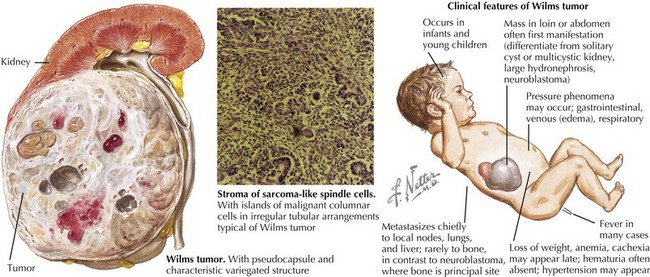


Wilms’ tumor is an embryonic solid tumor derived from undifferentiated nephroblasts in the kidney. Thus, the persistence of embryonic renal tissue (nephrogenic rests) is often seen in kidneys resected for Wilms’ tumor. The presence of multiple, diffuse nephrogenic rests is known as nephroblastomatosis, a premalignant condition prone to transforming into Wilms’ tumor. Not all nephrogenic rests transform into Wilms’ tumor; they may also regress or differentiate into mature kidney tissue. Therefore, children with diffuse nephroblastomatosis or a genetic predisposition for Wilms’ are at highest risk of nephrogenic rests progressing to malignant Wilms’ tumor.


Certain genetic syndromes are associated with the development of Wilms’ tumor. Examples include overgrowth syndromes such as Beckwith-Wiedemann (organomegaly, neonatal hypoglycemia, macroglossia, and omphalocele; also associated with hepatoblastoma) and Perlman’s (distinct facial features, macrosomia, genitourinary abnormalities and polyhydramnios), Denys-Drash (genital anomalies and nephropathy), and WAGR (Wilms’ tumor, aniridia, genitourinary anomalies, and mental retardation) syndromes. These patients have germline genetic abnormalities, specifically at chromosome 11, that alter the genes responsible for growth and renal genesis.


The 11p locus has been shown to play an important role in Wilms’ tumor pathogenesis. The WT1 gene, thought to be a tumor suppressor gene, is located at 11p13. Deletions of this region are associated with WAGR syndrome, and mutations of this gene are associated with Denys-Drash syndrome. Loss of heterozygosity or loss of imprinting at the WT2 gene, located at chromosome 11p15, has also been identified in patients with overgrowth syndromes such as Beckwith-Wiedemann and Perlman syndromes.


Specific genetic abnormalities that are exclusive to the tumor alone seem to be associated with a more aggressive form of Wilms’ tumor. The combined loss of heterozygosity of chromosomes 16q and 1p in Wilms’ tumors has been associated with a poorer prognosis in lower stage tumors. More recently, an X chromosome gene mutation, WTX, seen in one-third of sporadic Wilms’ tumors, has been described.


Although most Wilms’ tumors fall into the category of favorable histology, a subset of tumors can be categorized as diffusely “anaplastic,” having irregular mitoses and hyperchromatic cells with large nuclei throughout, which carries a poorer prognosis. No specific genetic abnormalities have been determined thus far for this aggressive subtype of Wilms’ tumor.


Rare Renal Tumors


Clear cell sarcoma of the kidney (CCSK) is the second most common malignant kidney tumor of childhood and accounts for about 2% to 4% of primary pediatric renal tumors. The majority of patients are younger than 5 years old at the time of diagnosis, and classic pathology shows nests of cells separated by septae. The majority of tumors also demonstrate variant patterns, which may resemble Wilms’ tumor and make it difficult to differentiate CCSK from other renal tumors.


Rhabdoid tumor of the kidney (RTK) is a very aggressive renal tumor that is most often detected in infants but is not exclusive to infancy. RTK, like its central nervous system homologue, atypical teratoid rhabdoid tumor (ATRT) of the brain, carries a specific deletion of the INI1 gene located at 22q11-2 that is a germline mutation in 20% of cases and can be used to confirm the diagnosis.


Renal cell carcinoma (RCC) is the most common form of adult renal cancer; however, pediatric RCC accounts for only 1% of all RCC and 5% to 6% of all pediatric renal tumors. Pediatric RCC seem to have a distinct tumor from adult RCC, with a genetic translocation involving chromosome Xp11. Interestingly, RCC has been described in a number of patients with a history of neuroblastoma. Renal medullary carcinoma (RMC), a subset of RCC, is extremely rare and is seen almost exclusively in young adolescent patients with sickle cell trait; the etiology and rationale for the association is unclear. Patients with RMC generally present in the second to third decade of life with advanced disease and have a very poor prognosis.


Cystic nephroma, on the other hand, is a rare and benign tumor of the kidney that is associated with a very favorable prognosis after surgical resection alone.


A kidney tumor noted on prenatal ultrasonography or in the first 6 months of life is almost always a congenital mesoblastic nephroma (CMN). CMN occurs in one in 27,500 live births and is the most common benign renal tumor of infancy. Because of the differential diagnosis of an infant renal tumor including malignant Wilms’ tumor and RTK, a nephrectomy is often performed to diagnose the lesion. The cellular variant of CMN is more aggressive with a 20% chance of local recurrence and shares the t(12;15)(p13;q26) gene fusion product ETV6-NTRK3 that is also found in infantile fibrosarcoma.


Clinical Presentation


The clinical presentation for patients with a renal tumor often includes abdominal distension, hematuria, hypertension, and a palpable abdominal mass. Symptoms may also be generalized and include fevers, unintentional weight loss, or night sweats.


Stay updated, free articles. Join our Telegram channel

Jun 19, 2016 | Posted by in PEDIATRICS | Comments Off on Renal Neoplasms

Full access? Get Clinical Tree

Get Clinical Tree app for offline access