Chapter 401 Pulmonary Embolism, Infarction, and Hemorrhage
401.1 Pulmonary Embolus and Infarction
Venous thromboembolic disease (VTE) is well described in children and adolescents with or without risk factors (Table 401-1). Improvements in therapeutics for childhood illnesses and increased survival with chronic illness may contribute to the larger number of children presenting with thromboembolic events, which can be a significant source of morbidity and mortality.
Table 401-1 RISK FACTORS FOR PULMONARY EMBOLISM
ENVIRONMENTAL
WOMEN’S HEALTH
MEDICAL ILLNESS
SURGICAL
THROMBOPHILIA
NONTHROMBOTIC
Modified from Goldhaber SZ: Pulmonary embolism, Lancet 363:1295–1305, 2004.
Etiology
Commonly appreciated risk factors for thromboembolic disease in adults include immobility, malignancy, pregnancy, infection, and hypercoagulability; up to 20% of adults with this disorder may have no identifiable risk factor (see Table 401-1). Children with deep venous thrombosis (DVT) and pulmonary embolism (PE) are much more likely to have 1 or more identifiable conditions or circumstances placing them at risk. In a large Canadian registry, 96% of pediatric patients were found to have 1 risk factor and 90% had 2 or more risk factors.
Children with malignancies are also at considerable risk. The risk of PE is more significant in children with solid rather than hematologic malignancies. PE has been described in children with Wilms tumor (tumor embolism) as well as leukemia. A child with malignancy may have numerous risk factors related to the primary disease process and the therapeutic interventions. Infection from chronic immunosuppression may interact with hypercoagulability of malignancy and chemotherapeutic effects on the endothelium.
In the neonatal period, thromboembolic disease and PE are often related to indwelling catheters used for parenteral nutrition and medication delivery. Emboli in neonates may occasionally reflect maternal risk factors, such as diabetes and toxemia of pregnancy. Infants with congenitally acquired homozygous deficiencies of antithrombin, protein C, and protein S are also likely to present with thromboembolic disease in the neonatal period.
Prothrombotic disease can also manifest in older infants and children. Disease can be congenital or acquired; DVT/PE may be the initial presentation. Factor V Leiden mutation (Chapter 472), hyperhomocysteinemia (Chapter 79.3), prothrombin 20210A mutation (Chapter 472), anticardiolipin antibody, and elevated values of lipoprotein A have all been linked to thromboembolic disease. Children with sickle cell disease are also at high risk for pulmonary embolus and infarction. Acquired prothrombotic disease is represented by nephrotic syndrome (Chapter 521) and antiphospholipid antibody syndrome. From one quarter to one half of children with systemic lupus erythematosus (Chapter 152) have thromboembolic disease.
Septic emboli are rare in children but may be caused by osteomyelitis, cellulitis, urinary tract infection, jugular vein or umbilical thrombophlebitis, and right-sided endocarditis.
Epidemiology
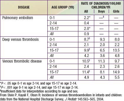
Younger age appears to be somewhat protective in thromboembolic disease. The DVT incidence in 1 study of hospitalized children was 5.3/10,000 admissions. A study that analyzed data from 1979 through 2001 found 0.9 cases of PE per 100,000 children per yr, 4.2 cases of DVT per 100,000 children per yr, and 4.9 cases of VTE per 100,000 children per yr (Table 401-2).
Pediatric autopsy reviews have estimated the incidence of thromboembolic disease in children as between 1% and 4%. Not all of these embolic findings were clinically significant. Thromboembolic pulmonary disease is often unrecognized, and antemortem studies may underestimate the true incidence. Pediatric deaths from isolated pulmonary emboli are rare. Most thromboemboli are related to central venous catheters. The source of the emboli may be lower or upper extremity veins as well as the pelvis and right heart. The most common location for an embolus unassociated with the presence of an indwelling venous catheter is the lower extremity.
Pathophysiology
Favorable conditions for thrombus formation include injury to the vessel endothelium, hemostasis, and hypercoagulability. Once an embolus develops and travels to the pulmonary circulation, symptoms of presentation are largely attributable to unequal ventilation and perfusion. The occlusion of the involved vessel prevents perfusion of distal alveolar units, thereby creating an increase in dead space and hypoxia with an elevated alveolar-arterial oxygen tension difference, or a-AO2 (PAO2 – PaO2). Most patients are hypocarbic secondary to hyperventilation, which often persists even when oxygenation is optimized. Abnormalities of oxygenation and ventilation are likely to be less significant in the pediatric population, possibly owing to less underlying cardiopulmonary disease and greater reserve. The vascular supply to lung tissue is abundant, and pulmonary infarction is unusual with pulmonary embolus but may result from distal arterial occlusion and alveolar hemorrhage. In rare instances of death from massive pulmonary embolus, marked increases in pulmonary vascular resistance and heart failure are usually present. Most of these severe outcomes are expectedly found in those with pre-existing cardiopulmonary disease.

Full access? Get Clinical Tree