Chapter 31 Puberty and Disorders of Pubertal development
This part of Essentials (Chapters 31 through 36) deals with the normal and abnormal hormonal influences on the female reproductive system. The sequence of events is an excellent example of the “Life-Course Perspective” for women’s health and health care introduced in Chapter 1. This series of events begins with endocrine changes in the fetus, the neonate, and then into childhood and pubertal development, then is followed by the early reproductive years, continuing on through the female climacteric, over the life course of a woman’s reproductive years. This section of the book concludes with a chapter on other common disorders that are influenced by normal and abnormal hormonal changes during the menstrual cycle.
The onset of pubertal changes is determined primarily by genetic factors, including race, and is also influenced by geographic location (girls in metropolitan areas, at altitudes near sea level, or at latitudes close to the equator tend to begin puberty at an earlier age) and nutritional status (obese children have an earlier onset of puberty, and those who are malnourished or who have chronic illnesses associated with weight loss have a later onset of menses). Excessive exercise relative to the caloric intake can also delay the onset of puberty. It has been proposed that an “invariant mean weight” of 48 kg (106 lb) is essential for the initiation of menarche in healthy girls and that leptin, a peptide secreted by adipose tissue, may be the link between weight and initiation of menarche. Psychological factors, severe neurotic or psychotic disorders, and chronic isolation may interfere with the normal onset of puberty through a mechanism similar to adult hypothalamic amenorrhea.
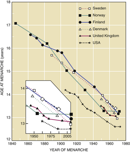
In the United States and Western Europe, a decrease in the age of menarche (age at first menses) was noted between 1840 and 1970. This trend has plateaued in the past 30 years (Figure 31-1). Presently, the mean age of menarche is about 12.4 years in the United States.

FIGURE 31-1 Decreasing age at menarche, 1840 to 1978, with inset from 1950 to 2008, indicating a leveling off (about 12.4 years in the United States) since 1975.
(Adapted from Styne DM, Grumbach MM: Disorders of puberty in the male and female. In Yen SSC, Jaffe RB, Barbieri RL [eds]: Reproductive Endocrinology: Physiology, Pathophysiology, and Clinical Management, 4th ed. Philadelphia, WB Saunders, 1999.)
Endocrinologic Changes of Puberty
FETAL AND NEWBORN PERIOD
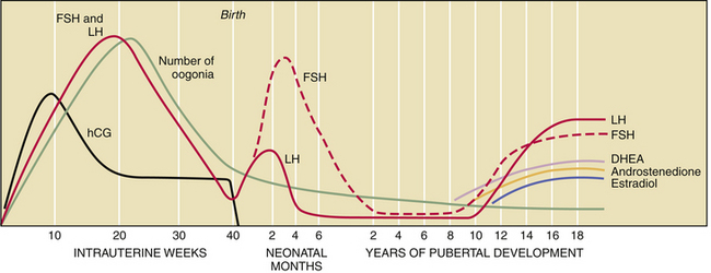
The fetal hypothalamic-pituitary-gonadal axis is capable of producing adult levels of gonadotropins and sex steroids. By 20 weeks’ gestation, levels of gonadotropins—follicle-stimulating hormone (FSH) and luteinizing hormone (LH)—rise dramatically in both male and female fetuses (Figure 31-2). The female fetus acquires the lifetime peak number of oocytes by mid-gestation, and she experiences a brief period of follicular maturation and sex steroid production in response to elevated gonadotropin levels in utero. This transient increase in serum estradiol (a sex steroid) acts on the fetal hypothalamic-pituitary unit, resulting in a reduction of gonadotropin secretion (negative feedback effect), which in turn reduces estradiol production. This indicates that the inhibitory effect of sex steroids on gonadotropin release is operative before birth.

FIGURE 31-2 Changes in the concentration of gonadotropins (LH and FSH), sex steroids (DHEA, androstenedione, and estradiol), and the number of oogonia throughout fetal life and pubertal development. hCG, human chorionic gonadotropin; LH, luteinizing hormone; FSH, follicle-stimulating hormone; DHEA, dehydroepiandrosterone.
(Adapted from Speroff L, Fritz M: Neuroendocrinology. In Speroff L, Fritz M [eds]: Clinical Gynecologic Endocrinology and Infertility, 7th ed. Baltimore, Williams & Wilkins, 2005.)
In both male and female fetuses, serum estradiol is primarily of maternal and placental origin. With birth and the acute loss of maternal and placental sex steroids, the negative feedback action on the hypothalamic-pituitary axis is lost, and gonadotropins are once again released from the pituitary gland, reaching adult or near adult concentrations in the early neonatal period. In the female infant, peak serum levels of gonadotropins are generally seen by 3 months of age and then slowly decline until a nadir is reached by the age of 4 years. In contrast to gonadotropin levels, sex steroid concentrations decrease rapidly to prepubertal values within 1 week after birth and remain low until the onset of puberty.
CHILDHOOD
The hypothalamic-pituitary-gonadal axis in the young child is suppressed between the ages of 4 and 10 years. The hypothalamic-pituitary system regulating gonadotropin release has been termed the gonadostat. Low levels of gonadotropins and sex steroids during this prepubertal period are a function of two mechanisms: maximal sensitivity of the gonadostat to the negative feedback effect of the low, circulating levels of estradiol present in prepubertal children, and intrinsic central nervous system inhibition of hypothalamic gonadotropin-releasing hormone (GnRH) secretion. These mechanisms occur independent of the presence of functional gonadal tissue. This is clearly demonstrated in children with gonadal dysgenesis. Agonadal children display elevated gonadotropin concentrations during the first 2 to 4 years of life, followed by a decline in circulating FSH and LH levels by 6 to 8 years of age. By 10 to 12 years of age, gonadotropin concentrations spontaneously rise once again, eventually achieving castration levels. This pattern of gonadotropin secretion in early childhood is similar to that of children with normal gonadal function. These data suggest that an intrinsic central nervous system regulator of GnRH release is the principal inhibitor of gonadotropin secretion from 4 years of age until the peripubertal period.
LATE PREPUBERTAL PERIOD
In general, androgen production and differentiation by the zona reticularis of the adrenal cortex are the initial endocrine changes associated with puberty. Serum concentrations of dehydroepiandrosterone (DHEA), dehydroepiandrosterone sulfate (DHEA-S), and androstenedione rise between the ages of 8 and 11 years. This rise in adrenal androgens induces the growth of both axillary and pubic hair and is known as adrenarche or pubarche. This increase in adrenal androgen production occurs independent of gonadotropin secretion or gonadal steroid levels, and the mechanism of its initiation is not understood at this time. Recent studies indicate that girls who undergo premature pubarche are more likely to develop polycystic ovary syndrome (PCOS) as adults.
PUBERTAL ONSET
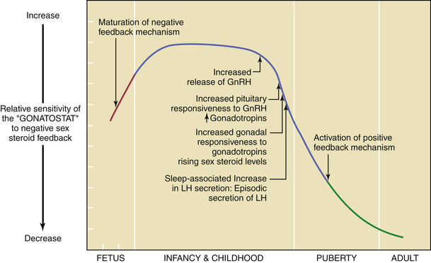
By about the 11th year of life, there is a gradual loss of sensitivity by the gonadostat to the negative feedback of sex steroids (Figure 31-3). As a consequence of this reduced negative feedback effect, GnRH pulses (with their mirroring pulses of FSH and LH) increase in amplitude and frequency. The factors that reduce the sensitivity of the gonadostat are incompletely understood. Some studies indicate that a rise in the concentration of leptin, a hormone produced by adipocytes (fat cells) that mediates appetite satiety, precedes and is necessary for this change. This, in turn, supports the connection between minimum weight or total body fat and the onset of puberty. A further decrease in sensitivity of the gonadostat, combined with the loss of intrinsic central nervous system inhibition of hypothalamic GnRH release, is heralded by sleep-associated increases in GnRH secretion. This nocturnal-dominant pattern gradually shifts into an adult-type secretory pattern, with GnRH pulses occurring every 90 to 120 minutes throughout the 24-hour day.

FIGURE 31-3 Changes in set point of the hypothalamic-pituitary unit (gonadostat) (solid lines) and the maturation of the negative and positive feedback mechanisms from fetal life to adulthood in relation to the normal changes of puberty. This figure does not illustrate the change in the sex steroid–independent intrinsic central nervous system inhibitory mechanism that is observed from late infancy to puberty. GnRH, gonadotropin-releasing hormone; LH, luteinizing hormone.
(Adapted from Styne DM, Grumbach MM: Disorders of puberty in the male and female. In Yen SSC, Jaffe RB [eds]: Reproductive Endocrinology: Physiology, Pathophysiology, and Clinical Management, 2nd ed. Philadelphia, WB Saunders, 1991.)
The increase in gonadotropin release promotes ovarian follicular maturation and sex steroid production, which induces the development of secondary sexual characteristics. By mid to late puberty, maturation of the positive-feedback mechanism of estradiol on LH release from the anterior pituitary gland is complete, and ovulatory cycles are established.
Somatic Changes of Puberty
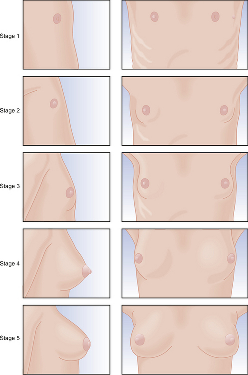
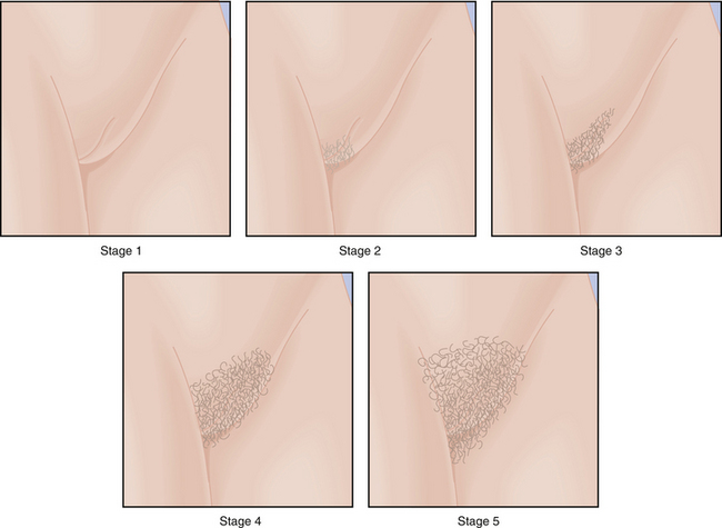
Physical changes of puberty involve the development of secondary sexual characteristics and the acceleration of linear growth (gain in height). The classification of breast and pubic hair development by Marshall and Tanner is employed for descriptive and diagnostic purposes (Figures 31-4 and 31-5).

FIGURE 31-4 Stages of breast development as defined by Marshall and Tanner. Stage 1: Preadolescent; elevation of papilla only. Stage 2: Breast bud stage; elevation of breast and papilla as a small mound with enlargement of the areolar region. Stage 3: Further enlargement of breast and areola without separation of their contours. Stage 4: Projection of areola and papilla to form a secondary mound above the level of the breast. Stage 5: Mature stage; projection of papilla only, resulting from recession of the areola to the general contour of the breast.
(Adapted from Marshall WA, Tanner JM: Variations in pattern of pubertal changes in girls. Arch Dis Child 44:291, 1969.)

FIGURE 31-5 Stage of female pubic hair development according to Marshall and Tanner. Stage 1: Preadolescent; absence of pubic hair. Stage 2: Sparse hair along the labia; hair downy with slight pigment. Stage 3: Hair spreads sparsely over the junction of the pubes; hair is darker and coarser. Stage 4: Adult-type hair; there is no spread to the medial surface of the thighs. Stage 5: Adult-type hair with spread to the medial thighs assuming an inverted triangle pattern.

Stay updated, free articles. Join our Telegram channel

Full access? Get Clinical Tree

