Chapter 12 Prolapse and Urogynaecology
Retroversion of the uterus
An alteration from the usual anteverted position of the uterus often with a change in the curve of the uterine axis. Most of the so-called displacements are merely variations of the normal and are of little clinical significance.
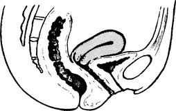
Anteverted Uterus
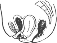
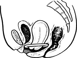
The uterus is approximately at right angles to the vagina and has a slight forward curve.
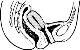
Retroversion
The long axis of the uterus is directed backwards. The uterus is displaced backwards and this is simply a physiological variation for the vast majority of women. It occurs in approximately 20–30% of women.
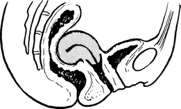
Retroflexion
This is a variation of retroversion but the uterus is curved backwards. The cervix may remain in the normal position but is usually positioned as in retroversion.
Causes of Displacement
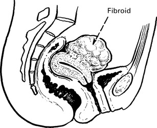
Displacement may be due to the presence of some other condition such as a cyst or fibroid or endometriosis.
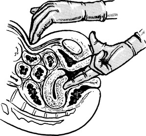
Diagnosis is by bimanual palpation. The vaginal hand palpates a mass in the Pouch of Douglas, the abdominal hand detects the absence of a uterine corpus in the expected place. The possibility of pelvic pathology should be considered and ultrasound may be used to confirm the clinical findings.
Symptoms and treatment of displacement
Consequences of Uncomplicated Displacement
Usually none. However, if there is pelvic pathology present, the woman may complain of deep dyspareunia. If such pain occurs, it can easily be reproduced by pressing with the examining fingers. It is quite possible for the patient to present with a complaint of dyspareunia and to have a retroverted uterus which has nothing to do with her complaint.
Uterovaginal prolapse
Herniation of the genital tract through the pelvic diaphragm.
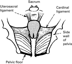
The uterus and vagina are held in the pelvis by the cardinal and uterosacral ligaments and by the pelvic floor musculature, mainly the levatores ani.
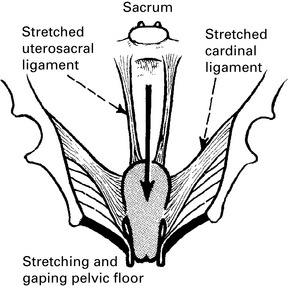
When these ligaments and muscles become ineffective, the uterus and vagina descend (prolapse) through the gap between the muscles.
The causes of prolapse are the following:
The incidence of this condition in the United Kingdom has been greatly reduced because of smaller families and higher caesarean section rates.
Uterovaginal prolapse
The uterus gradually descends in the axis of the vagina taking the vaginal wall with it. It may present clinically at any level, but is usually classified as one of three degrees.
First degree: cervix still inside vagina.
Second degree: the cervix appears at the level of the introitus.
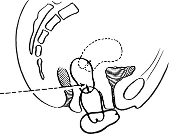
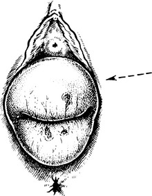
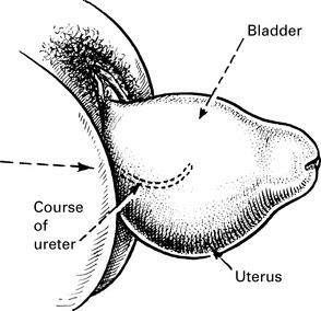
Third degree: complete prolapse. In the picture, the uterus is retroflexed and the outline of bladder can be seen. There may be a rectal prolapse as well. This is sometimes called complete procidentia. The vaginal mucosa is non-keratinised and the tissues can become very dry and ulcerated with a 3rd degree uterine prolapse. Bleeding and infection can also occur.
Vaginal prolapse
The prolapse involves the vaginal walls and the related viscera. Prolapse of several sites may co-exist and a uterine prolapse may also be present.
Anterior prolapse
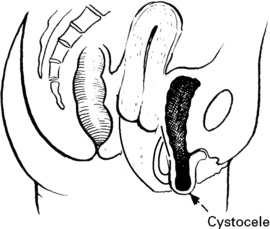
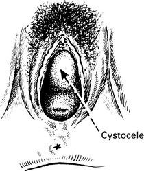
When the upper part of the anterior wall prolapses, there is an underlying failure of the investing fascia, and the bladder base also descends. This is called a cystocele.
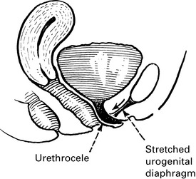
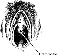
Sometimes the lower part of the vaginal wall prolapses and the urethra also descends. This is called a urethrocele.
Vaginal prolapse
Prolapse of the posterior wall
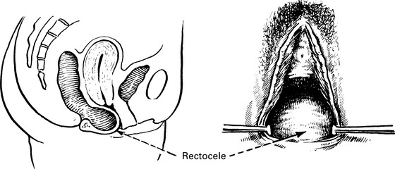
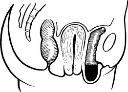
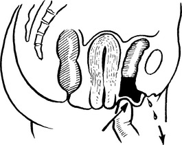
If the prolapse is at the level of the middle third of the vagina, the rectovaginal septum is often involved and rectum prolapses with vaginal wall. This is called a rectocele. If the lowest part of the vagina prolapses, the perineal body is involved rather than the rectum.
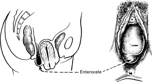
If the upper part of the posterior vaginal wall prolapses, the Pouch of Douglas is elongated and small bowel or omentum may descend. This is called an enterocele. Enterocele is often associated with uterine prolapse, as in the picture.
Where the uterus has been removed, prolapse of this region is known as a vault prolapse. This is a difficult condition to treat. The technique depends on suspending the vaginal vault from fixed ligaments within the pelvic and often requires insertion of a non-absorbable mesh.
Clinical features of prolapse
The onset may be gradual or quite sudden and is commoner after the menopause when the genital tract tissues begin to atrophy.
Differential diagnosis of prolapse
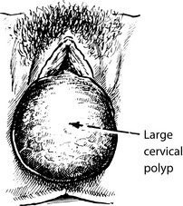
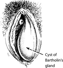
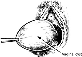
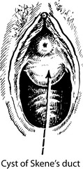
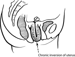
Prolapse should be confirmed by vaginal examination. The following conditions resemble prolapse on superficial examination; however, careful examination will detect the difference.
Pessary treatment
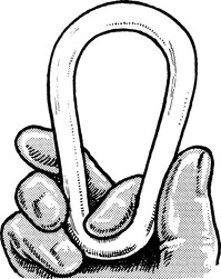
There are a number of different shapes of vaginal pessaries, the simplest to insert is a ring pessary. This is usually made of semi-rigid plastic and is inserted into the vagina so that the vaginal walls are stretched and they cannot prolapse through the introitus.
The pessary is compressed into a long ovoid shape, lubricated and gently pushed into the vagina, where it resumes its circular shape and takes up a position in the coronal plane. It must not be too tight; and correct fitting is learnt by experience. To an extent, pessary fitting is trial and error and the woman should be warned that the pessary may dislodge. A point of contact should be given so that she can be seen again without delay if this is the case.
Alternative-shaped pessaries
The ‘shelf’ pessary can be particularly useful for uterine prolapse but it is slightly less malleable and insertion can be more difficult.
Indications for pessary treatment
Plastic rings should be changed once or twice a year, and if it has been properly fitted, the patient will be unaware of its presence in her vagina even during coitus. An ill-fitting pessary is ineffective and may cause dyspareunia and discomfort. If it is too tight or left too long, ulceration of the vaginal wall will occur, and malignant change has been reported. Vaginal oestriol cream or vaginal oestradiol tablets (0.025 mg) help prevent atrophic vaginitis.

Full access? Get Clinical Tree