Fig. 7.1
This is the classical image of PCOS with an enlarged ovary containing an increased number of small follicles around the periphery of the cortex, resembling a string of pearls, along with a bright echogenic stroma
The differential diagnosis should include other causes of hyperandrogenism and oligomenorrhea such as nonclassical congenital adrenal hyperplasia (CAH) , hypothalamic hypogonadism , Cushing’s syndrome and disease, hyperprolactinemia, thyroid disease, acromegaly, androgen-secreting neoplasms of the ovary or adrenal gland, and exogenous steroid use. The ontogeny of androgen profiles has been described in a Nordic multicenter collaborative study. Women with PCOS also had elevated serum androgen levels after menopause. In the absence of high sensitivity and high specificity testosterone assays, the best predictive hormone was androstenedione [3].
7.3 Prevalence
The prevalence of PCOS is estimated to be 4–12% of reproductive-age women. The largest US study on PCOS prevalence was published in 1998 [4]. Out of 277 women included in the study, 4.0% had PCOS as defined by the 1990 NIH criteria. The prevalence was 4.7% for white women and 3.4% for black women. The inclusion of polycystic ovaries in the 2003 Rotterdam criteria calls for reevaluation of the prevalence of PCOS, as 21–23% of normal women have polycystic-appearing ovaries on ultrasound.
7.4 Clinical Case
7.4.1 Hyperandrogenism
Clinical manifestations of hyperandrogenemia include hirsutism , acne, and male pattern alopecia. Hirsutism is defined as the growth of coarse, pigmented hairs in androgen-dependent areas such as the face, chest, back, and lower abdomen. Approximately 80% of hirsute patients will have PCOS [5]. The modified Ferriman–Gallwey scoring system can be used for clinical assessment of hirsutism . This system, which was originally used in the United Kingdom for a population of presumably Caucasian women, scores hair growth in nine body areas from 0 (absence of terminal hairs) to 4 (extensive terminal hair growth) [6]. Other hyperandrogenic manifestations commonly found in PCOS patients include acne and alopecia [7, 8]. Acne is a result of androgen stimulation of the pilosebaceous unit with increased skin oiliness [7].
7.4.2 Obesity
Obesity is very common in PCOS, with the android pattern present in approximately 44% of women with PCOS [9]. This central obesity is more characteristic of PCOS, as these patients have an increased waist-to-hip ratio compared to obese women without PCOS [10]. Hyperinsulinemia may stimulate central adiposity, which, in turn, exacerbates underlying or latent insulin resistance [11].
7.4.3 Insulin Resistance, Diabetes, and Acanthosis Nigricans
Insulin resistance and diabetes are important health concerns commonly seen in association with polycystic ovarian syndrome and will be discussed at length later in this chapter. Acanthosis nigricans is a dermatological condition of hyperkeratosis and increased skin pigmentation with raised, symmetrical, darkened, velvety plaques that commonly appear on the nape of the neck. It can also be found in the axilla, groin, and other intertriginous areas of the body. Elevated insulin has a mitogenic effect on basal cells of the epidermis, making acanthosis nigricans a relatively specific clinical marker of insulin resistance [12].
7.4.4 Irregular Menses and Infertility
Some of the menstrual abnormalities seen with chronic anovulation include secondary amenorrhea, oligomenorrhea , and dysfunctional uterine bleeding. Menarche typically begins at a normal or early age, but the menstrual irregularities often seen in adolescents may never resolve for the PCOS patient. The irregular menses may be masked in PCOS patients if they are on oral contraceptives. PCOS is the most common cause of anovulatory infertility, which often serves as the impetus for the patient to seek medical attention. Patients may report false-positive urinary ovulation predictor tests due to chronic elevation in luteinizing hormone (LH).
7.4.5 Miscarriage
The risk of a first-trimester spontaneous abortion is reported to be significantly higher for patients with PCOS. The spontaneous abortion rate in PCOS is reported to be 30% [13]. In comparison, retrospective studies find the risk of spontaneous abortion to be 5–14% for normal women [14, 15]. Of patients with recurrent miscarriage, 36–82% have polycystic ovaries [13, 16, 17].
Several explanations have been offered. For example, Homburg et al. demonstrated that high concentrations of LH during the follicular phase in women with polycystic ovaries have a deleterious effect on rates of conception and are associated with early pregnancy loss [18].
7.5 Pathogenesis
7.5.1 Altered Gonadotropin Secretion
One of the well-described features of PCOS is an increase in LH and relative decrease in follicle-stimulating hormone (FSH) [19]. The relative decrease in FSH is the chief cause of anovulation. The pulsatile secretion of LH from the pituitary is increased in amplitude and frequency [20]. In addition, the pituitary has a greater LH response to gonadotropin-releasing hormone (GnRH) compared with normal women [20, 21].
The pulsatile secretion of GnRH cannot be studied in humans, so it must be inferred by detecting peripheral LH patterns. A study of PCOS women by Berga et al. found increased pulse frequency and amplitude for LH and α (alpha)-subunit, providing evidence for aberrant increases in GnRH pulse frequency (◘ Fig. 7.2) [20]. Elevated LH is not caused by altered pituitary sensitivity to GnRH , as GnRH receptor blockade resulted in similar LH decreases in PCOS and normal women [22]. These findings suggest a derangement of the hypothalamic–pituitary axis , which appears to play a major role, because many of the cardinal features of PCOS can be traced to alterations in gonadotropins .


Fig. 7.2
Twenty-four hour concentration profiles of LH (top) and α(alpha)-subunit (bottom) in an eumenorrheic woman (EW) (left), studied in the follicular phase (day 2) and in a woman with hyperandrogenic anovulation (HAA) /PCOS (right). Reprinted with permission from Berga S, Guzick D, Winters S. Increased luteinizing hormone and alpha-subunit secretion in women with hyperandrogenic anovulation. J Clin Endocrinol Metab 1993; 77(4):895–901. Copyright 1993, The Endocrine Society
7.5.2 Neuroanatomical Considerations
The GnRH pulse generator refers to the synchronized pulsatile secretion of GnRH from neurons that are widely distributed in the medial basal hypothalamus. Knobil and associates conducted experiments with the Rhesus monkey to establish that the GnRH system exhibits rhythmic electrical behavior in the arcuate nucleus of the medial basal hypothalamus [23]. There was remarkable synchrony between pulses of GnRH in the portal blood and LH pulses in peripheral blood. This phenomenon was later studied in isolated human medial basal hypothalamus, where GnRH pulses were found to occur at a frequency of 60–100 min [24].
The secretion of GnRH into the portal vasculature also appears to be regulated by dynamic remodeling of GnRH neurovascular junctions. Morphological plasticity of the median eminence during the menstrual cycle has been demonstrated, where the maximal number of GnRH neuro-vasculature junctions are found during the LH surge [25].
7.5.3 GnRH Neuroregulation in PCOS
The GnRH pulse generator in PCOS patients is intrinsically faster, and the frequency is less likely to be suppressed with continuous estrogen and progesterone treatment [26].
Increased central adrenergic tone has been implicated as a cause of the aberrations of GnRH and gonadotropin secretion in PCOS. One possible mechanism is the increase in local blood flow and permeability of the portal vascular system, permitting the entry of increased amounts of GnRH [27]. Dopamine injection into the third ventricle led to a rapid increase in GnRH and prolactin inhibitory factor in portal blood, suggesting dopamine-mediated regulation of GnRH and prolactin inhibitory factor [28]. The identification of β(beta)-1-adrenergic and D1-dopaminergic receptors on GT-1 GnRH neurons provides a mechanism by which norepinephrine and dopamine could regulate gonadotropin release via direct synapses on GnRH neurons [29].
The role of insulin-like growth factor 1 (IGF-1) in modulation of GnRH cells has also been investigated. IGF-1 regulates growth, differentiation, survival, and reproductive function. The IGF receptor is a tyrosine kinase receptor located in the periphery and CNS, including the median eminence [30]. In PCOS women, an increased ratio of IGF-1 to their binding proteins correlated significantly with increased concentrations of circulating LH [21]. These findings suggest that IGF-1 can modulate GnRH neurons by inducing gene expression, resulting in more circulating LH .
7.5.4 Hyperandrogenemia
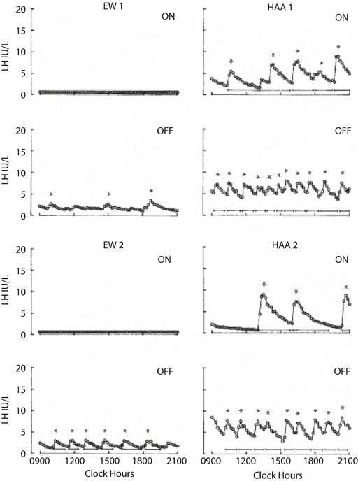
Circulating androgens are elevated in PCOS, with contributions from the ovary and adrenal glands. The elevated androgens can only be partially suppressed with combined oral contraceptive (COC) therapy . Daniels and Berga treated PCOS women with 3 weeks of COCs and found that androstenedione levels remained significantly higher compared to treated controls [26]. Pulse frequency of LH was suppressed in both PCOS women and controls, but the frequency remained significantly higher in PCOS patients (◘ Fig. 7.3). This suggests there is reduced sensitivity of the GnRH pulse generator to suppression by sex steroids. The authors also suggest that GnRH drive in PCOS women may be intrinsically and irreversibly faster than in eumenorrheic women.


Fig. 7.3
Representative 12-h pulse patterns in two women with polycystic ovary syndrome/HAA are shown on the right side and those from eumenorrheic women on the left. “ON” means subjects were studied on day 21 of a combined oral contraceptive containing 35 μg of ethinyl estradiol and 1 mg of norethindrone. “OFF” refers to day 7 following cessation of the combined oral contraceptive. Reprinted with permission from Daniels T, Berga S. Resistance of gonadotropin releasing hormone drive to sex steroid-induced suppression in hyperandrogenic anovulation. J Clin Endocrinol Metab 1997; 82(12):4179–4183. Copyright 1997, The Endocrine Society
7.5.5 Theca Cell Function
Ovarian hyperandrogenism is driven by LH acting on theca cells, and the effect is amplified by the increased sensitivity of PCOS theca cells to LH [31]. Hyperandrogenism may also result from dysregulation of the androgen-producing enzyme P450c17, which has 17 α (alpha)-hydroxylase and 17,20-lyase activities. In contrast, in vivo studies do not find significant increases in androgen secretion in women with PCOS or normal women, despite considerable increases in insulin levels. A role for insulin is strongly suggested by the observation that reduction of hyperinsulinemia is associated with decreases of serum androgens. Treatment of PCOS patients with metformin, which reduces hepatic glucose production and secondarily lowers insulin, has been shown to decrease levels of testosterone, DHEAS , and androstenedione [32].
7.5.6 Adrenal Function
Excess adrenal androgen production is seen in PCOS women, with a 48–64% increase in DHEAS and 11β (beta)-hydroxyandrostenedione. The underlying cause of elevated adrenal androgens is yet to be elucidated, but PCOS women do not have increased adrenocorticotropic hormone (ACTH ) levels [33]. Increased adrenal androgen production in PCOS is likely caused by either altered adrenal responsiveness to ACTH or abnormal adrenal stimulation by factors other than ACTH.
7.5.7 Anovulation
The cause of anovulation in PCOS patients has yet to be clarified. However, several observations in granulosa function have been described that may give insight into this process.
7.5.8 Granulosa Cell Function
FSH levels are characteristically low in PCOS women, resulting in arrested follicular development. Insufficient granulosa cell aromatase activity was the basis of earlier studies that tried to explain poor follicular development, as follicular fluid estradiol concentrations were thought to be low. To the contrary, more recent studies found that PCOS granulosa cells are hyperresponsive to FSH in vitro, and estradiol concentrations from PCO follicles and normal follicles are no different [34]. A dose response study in PCOS women demonstrated a significantly greater capacity for estradiol production in response to recombinant human FSH compared with normal women [35]. The incremental response of serum estradiol was almost two times greater and considerably accelerated compared with that found in normal women .
7.5.9 Insulin Resistance
Although 50–70% of PCOS patients have insulin resistance [36], it is not one of the diagnostic criteria of PCOS. The topic deservedly receives much attention, as many of the clinical signs and symptoms of PCOS may be attributed to excess insulin exposure. The precise molecular basis for insulin resistance is unknown, but it appears to be a postreceptor defect [37]. There is tissue specificity of insulin resistance in PCOS: muscle and adipose tissue are resistant, while the ovaries, adrenals, liver, skin, and hair remain sensitive. The resistance to insulin in skeletal muscle and adipose tissue leads to a metabolic compromise of insulin function and glucose homeostasis, but there is preservation of the mitogenic and steroidogenic function in other tissues. The effect of hyperinsulinemia on the sensitive organs results in downstream effects seen in PCOS, such as hirsutism [5], acanthosis nigricans [12], obesity [11], stimulation of androgen synthesis, increase in bioavailable androgens via decreased sex hormone-binding globulin (SHBG ) [38], and, potentially, modulation of LH secretion.
In 1992, Hales and Barker proposed the concept that the environmental influence of undernutrition in early life increased the risk of type 2 diabetes in adulthood [39]. They discovered a relationship between low birth weight and type 2 diabetes in men from England. In the “thrifty phenotype hypothesis,” malnutrition serves as a fetal and infant insult that results in a state of nutritional thrift. The adaptations result in postnatal metabolic changes that prepare the individual for survival under poor nutritional conditions. The adaptations become detrimental when the postnatal environment changes to one of an overabundance of nutrients, resulting in obesity and diabetes.
Insulin resistance is a component of the World Health Organization (WHO) definition of the metabolic syndrome, which is a cluster of risk factors for cardiovascular disease [40]. The WHO defines the metabolic syndrome as the presence of glucose intolerance or insulin resistance, with at least two of the following: hypertension, dyslipidemia, obesity, and microalbuminuria. Women with PCOS are 4.4 times more likely to have the metabolic syndrome, so it becomes prudent to screen these patients, especially in those with insulin resistance [41].
Lipid abnormalities are also more prevalent in PCOS patients. There can be a significant increase in total cholesterol, LDL cholesterol, and triglycerides, and a decrease in HDL cholesterol compared to weight-matched controls [42]. The dyslipidemia, impaired glucose intolerance, central obesity, hyperandrogenism , and hypertension seen in PCOS patients greatly increase the risk for cardiovascular disease. Based on this risk profile, women with PCOS have a sevenfold increased risk of myocardial infarction [43].
7.5.10 Laboratory Evaluation
In addition to confirming elevations of androgens, the laboratory evaluation of PCOS should have the objective of excluding other causes of hyperandrogenic anovulation. Androgen-producing tumors of the ovary and adrenals must be excluded. The adrenal glands contribute 98% of circulating DHEAS , while both the ovaries and adrenals contribute equal amounts of circulating testosterone and androstenedione. If total testosterone is greater than 200 ng/dL or DHEAS is greater than 7000 ng/dL, MRI is warranted to identify the hormone-secreting lesion. Measuring 17 α (alpha)-hydroxyprogesterone will screen for 21-hydroxylase deficiency, the most common enzyme deficiency in nonclassical CAH. A 17-hydroxyprogesterone level of greater than 3 ng/mL is defined as elevated and should be followed by an ACTH stimulation test, using 250 μg of synthetic ACTH given intravenously following an overnight fast. A 1-h increase of 17 α (alpha)-hydroxyprogesterone of more than 10 ng/mL is indicative of an enzyme defect in 21-hydroxylase.
Cushing’s syndrome may masquerade as PCOS. Those who have additional signs of Cushing’s syndrome, such as a moon facies, buffalo hump, abdominal striae, easy bruising, and proximal myopathy, should undergo screening with a 24-h urinary-free cortisol. In the work-up for anovulation, exclusion of prolactinoma should be performed. It is not uncommon to detect mild elevations in prolactin levels in PCOS patients. Thyroid-stimulating hormone (TSH) should be evaluated. LH, FSH , and estradiol levels should be obtained to exclude hypothalamic amenorrhea or premature ovarian failure.
7.5.11 Diabetes Screen/Evaluation of Insulin Resistance
The 2003 Rotterdam PCOS consensus group recommends a 2-h oral glucose tolerance test (OGTT) for obese PCOS patients and nonobese PCOS patients with risk factors for insulin resistance, such as family history of diabetes [2]. Defining insulin resistance is difficult, because the concept is nebulous with no universally accepted diagnostic strategy. The WHO defines insulin resistance as the lowest quartile of measures of insulin sensitivity. Women with PCOS are at significantly increased risk for impaired glucose tolerance and type 2 diabetes compared to age-, weight-, and ethnicity-matched controls [44]. If either the fasting glucose is 126 mg/dL or more, or the 2-h level is 200 mg/dL or more, diabetes is detected and should be confirmed with a repeat test. Impaired fasting glucose is defined as a glucose level between 100 and 126 mg/dL. Impaired glucose tolerance is defined as a 2-h glucose level between 140 and 200 mg/dL. It is also reasonable to obtain a fasting lipid profile in women suspected of having risk factors for cardiovascular disease. HgbA1C has been recently advocated as an accurate screening tool in evaluation of insulin resistance and diabetes in women with PCOS [45].
Stay updated, free articles. Join our Telegram channel

Full access? Get Clinical Tree


